Quantitative use of tracer cells
- Summary
- Abstract
- Description
- Claims
- Application Information
AI Technical Summary
Problems solved by technology
Method used
Image

Examples
example 1
Interference by Lubricants
Objective:
[0032] The objective was to determine the effect of various lubricant formulations in the ThinPrep® 2000 System.
Design:
[0033] Samples were prepared in 20 ml (final volume) of PreservCyt® Solution that contained 360,000 CaSki cells and 40,000 pre-stained CaSki tracer cells. The “test” samples also contained 100 mg of lubricant. Three different lubricant formulations, designated “A”, “B”, and “C”, from the same manufacturer were examined. Test samples were prepared by adding 1 ml of 10% lubricant stock solutions (prepared in PreservCyt® Solution) to 19 ml of the cell mixture. Control samples, containing no lubricant, were prepared by adding 1 ml of PreservCyt® Solution to 19 ml of the cell mixture.
[0034] Slides were prepared from each sample using the ThinPrep® 2000 processor. After preparation, slides were exposed to 95% alcohol and xylene and cover-slipped without any further cell staining. Slides were then imaged on the ThinPrep® Imaging S...
example 2
Interaction of Preservative Formulation and Blood
Objective:
[0041] The objective was to determine the performance of the ThinPrep System for preparing slides from samples prepared in two different formulations of cell preservative and in the absence or presence of blood.
Experimental Design:
[0042] Samples were prepared in 20 ml (final volume) of either cell preservative “A” or “B” that contained 300,000 CaSki cells and 100,000 pre-stained CaSki tracer cells. 100 ul of human blood (citrate treated to prevent coagulation) was added to half of the samples. Slides were prepared from each sample using the ThinPrep® 2000 processor. After preparation, slides were exposed to 95% alcohol and xylene and cover-slipped without any further cell staining. Slides were then imaged on the ThinPrep® Imaging System and tracer cell counts obtained from the imaged data using the CellCount and Kaleido-Spot software programs.
[0043] Results:
TABLE 2BloodNoYesCell“A”20,880639Preservative“B”35,6194,851...
example 3
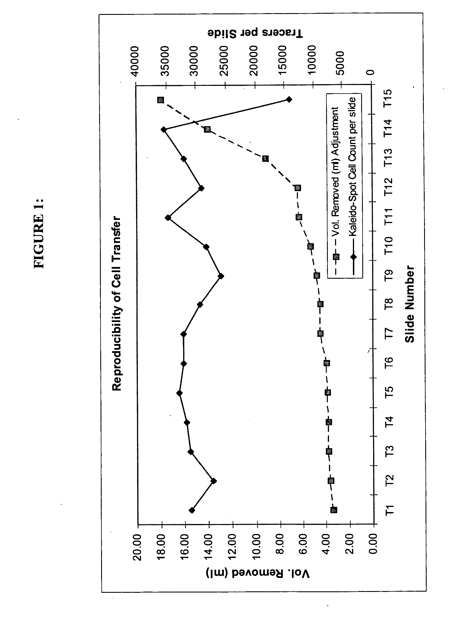
Reproducibility of Preparing Multiple Successive Slides
Objective:
[0046] The objective was to determine the consistency of slides prepared sequentially from the same specimen.
Experimental Design:
[0047] A 20 ml sample was prepared in PreservCyt® Solution. This sample was comprised of a pool of residual clinical specimens containing about 1,600,000 epithelial cells and 600,000 pre-stained CaSki tracer cells.
[0048] This 20 ml cell mixture was dispensed into a specimen vial and a slide prepared on the ThinPrep® 2000 processor. The volume remaining in the vial was measured and recorded. The volume necessary to produce the slide was calculated (20 ml minus the remaining volume) and this volume of PreservCyt® Solution was added to the vial to restore the sample volume to 20 ml.
[0049] A second slide was then produced on the ThinPrep® 2000 processor and the remaining volume measured and recorded. As before, the volume of sample removed by the processor was replaced with fresh PreservC...
PUM
 Abstract
Description
Claims
Application Information
 - R&D Engineer
- R&D Manager
- IP Professional
- Industry Leading Data Capabilities
- Powerful AI technology
- Patent DNA Extraction
Browse by: Latest US Patents, China's latest patents, Technical Efficacy Thesaurus, Application Domain, Technology Topic.
© 2024 PatSnap. All rights reserved.Legal|Privacy policy|Modern Slavery Act Transparency Statement|Sitemap