
Transvisceral neurostimulation mapping device and method

a mapping device and transvisceral technology, applied in the field of transvisceral neurostimulation mapping device and method, can solve the problem that the neurostimulation mapping tool lacks the ability to mark stimulation locations

Inactive Publication Date: 2007-06-28
SYNAPSE BIOMEDICAL INC
View PDF94 Cites 47 Cited by
  • Summary
  • Abstract
  • Description
  • Claims
  • Application Information

AI Technical Summary

Benefits of technology

[0017] The novel features of the invention are set forth with particularity in the appended claims. A better understanding of the features and advantages of the present invention will be obtained by reference to the following detailed description that sets forth illustrative embodiments, in which the principles of the invention are utilized, and the accompanying drawings of which:

Problems solved by technology

In addition, earlier neurostimulation mapping tools lacked the ability to mark stimulation locations, thus requiring the use of a separate marking tool.

Method used


Image

  • Transvisceral neurostimulation mapping device and method
  • Transvisceral neurostimulation mapping device and method
  • Transvisceral neurostimulation mapping device and method

Examples


example 1

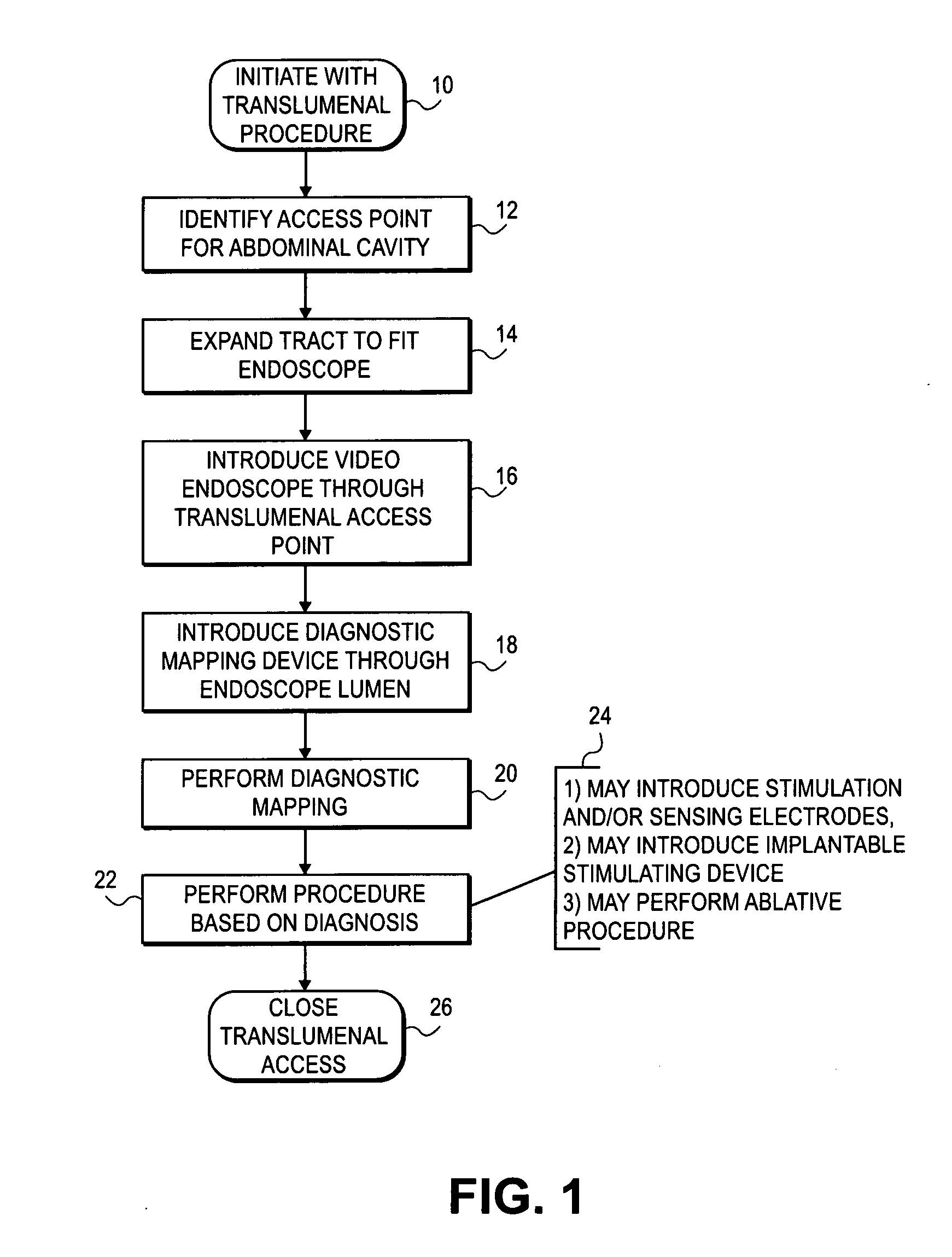
[0049] Methods: Pigs were anesthetized and transgastric peritoneal access with a flexible endoscope was obtained using a guidewire, needle knife cautery and balloon dilatation. The diaphragm was mapped to locate the motor point (where stimulation provides complete contraction of the diaphragm) with an endoscopic electrostimulation catheter. An intramuscular electrode was then placed at the motor point with a percutaneous needle. This was then attached to the diaphragm pacing system. The gastrotomy was managed with a gastrostomy tube.

[0050] Results: Four pigs were studied and the diaphragm could be mapped with the endoscopic mapping instrument to identify the motor point. In one animal, under trans-gastric endoscopic visualization a percutaneous electrode was placed into the motor point and the diaphragm could be paced in conjunction with mechanical ventilation.

[0051] Conclusion: These animal studies support the concept that transgastric mapping of the diaphragm and implantation of...

example 2

[0052] Methods: Four female pigs (25 kg) were sedated and a single channel gastroscope was passed transgastrically into the peritoneal cavity. Pneumoperitoneum was achieved via a pressure insulator through a percutaneous, intraperitoneal 14-gauge catheter. Three other pressures were recorded via separate catheters. First, a 14-gauge percutaneous catheter passed intraperitoneally measured true intra-abdominal pressure. The second transducer was a 14-gauge tube attached to the endoscope used to measure endoscope tip pressure. The third pressure transducer was connected to the biopsy channel port of the endoscope. The abdomen was insulated to a range (10-30 mmHg) of pressures, and simultaneous pressures were recorded from all pressure sensors.

[0053] Results: Pressure correlation curves were developed for all animals across all intraperitoneal pressures (mean error −4.25 to −1 mmHg). Endoscope tip pressures correlated with biopsy channel pressures (R2=0.99). Biopsy channel and endoscop...


PUM


Abstract

The invention provides a method and device for providing electrical stimulation to a patient's diaphragm (or other organ or tissue) including the steps of: introducing an endoscope transviscerally (e.g., transgastrically) into the patient's body cavity; delivering an electrode into the patient's body cavity through a lumen of the endoscope; applying suction to attach the electrode to a stimulation site on the diaphragm (or other organ or tissue); and delivering a stimulation pulse to the stimulation site. The stimulation may be repeated at multiple stimulation sites.

Description

CROSS-REFERENCE [0001] This application claims the benefit under 35 U.S.C. § 119 of U.S. Patent Application No. 60 / 597,440 filed Dec. 2, 2005, and which is incorporated herein by reference in its entirety.BACKGROUND OF THE INVENTION [0002] Electrodes are implanted into patients for a variety of purposes, such as to stimulate muscle movement and to provide pain relief. For example, U.S. Pat. Nos. 5,472,438 and 5,797,923 and U.S. Patent Appl. Publ. No. 2005 / 0021102 describe neurostimulation of a patient's diaphragm to assist the patient's breathing. [0003] Correct placement of stimulation electrodes helps achieve the best results. For example, optimal neurostimulation of a patient's diaphragm requires placement of the stimulation electrode or electrodes at or near phrenic nerve motor points. As described in U.S. Patent Appl. Publ. No. 2005 / 0021102, the desired stimulation electrode placement may be determined via a mapping procedure in which a mapping electrode is temporarily placed o...

Claims


Application Information

Patent Timeline
Patent Type & Authority Applications(United States)
IPC IPC(8): A61N1/00
CPCA61B1/2736A61B1/313A61B5/04001A61B5/05A61B5/4893A61B2017/306A61N1/05A61N1/0509A61N1/0517A61N1/0551A61N1/36007A61N1/3601A61B5/24A61B5/388
Inventor IGNAGNI, ANTHONY R.ONDERS, RAYMOND P.GELBKE, JAMES E.
Owner SYNAPSE BIOMEDICAL INC
Learn More