
Apparatus and method for harvesting and handling tissue samples for biopsy analysis

a biopsy analysis and tissue technology, applied in the field of tissue analysis, can solve the problems of difficult retrieval and processing, small sample size, and inability to determine the cell type of the tumor by a pathologis

Inactive Publication Date: 2007-07-19
BIOPATH AUTOMATION
View PDF99 Cites 117 Cited by
  • Summary
  • Abstract
  • Description
  • Claims
  • Application Information

AI Technical Summary

Benefits of technology

[0085] The inventive system disclosed herein has been designed to move the biopsy samples with the least amount of operator involvement, while maintaining at least the current standards for preparing slides. For instance, numerous methods are disclosed which take advantage of the invention's ability to capture tissue samples at the site of harvest, thus eliminating the steps of transferring the samples form one container to another. The inventive material goes on to encompass the entire record keeping, tissue processing and wax embedding procedures. Therefore, a larger number of samples can be processed by a fewer number of individuals. This is accomplished by utilizing the inventive tissue handling components, combined with automated machinery to transport tissue samples through the various stages creating a final slide for pathological examination.
's ability to capture tissue samples at the site of harvest, thus eliminating the steps of transferring the samples form one container to another. The inventive material goes on to encompass the entire record keeping, tissue processing and wax embedding procedures. Therefore, a larger number of samples can be processed by a fewer number of individuals. This is accomplished by utilizing the inventive tissue handling components, combined with automated machinery to transport tissue samples through the various stages creating a final slide for pathological examination.

Problems solved by technology

While radiologic tools are useful in detecting the presence of a tumor, the cell type of the tumor can only be determined by a pathologist's examination of a histologic or cytologic sample of the tumor.
In many cases, these samples are very small and difficult to retrieve and process.
Active needles have two basic problems, which are cost and complexity.
0.016″ does not provide a great deal of clearance for these elements and thus these prior art needles are inefficient.
Other methods of obtaining samples are also discussed in the literature and also have problems.
Some samples can be as small as a few cells, and extremely small samples can create problems.
These problems include loss of the sample, dehydration of the sample, and contamination of the sample during harvesting, storage and transport.
Still further, as will be more evident from the following discussion, small samples are extremely difficult and time consuming to process in the laboratory.
Prior art devices and methods account for collection of effluent only and do not provide devices and methods for trapping tissue specimens.
The prior art collects effluent, but does not provide devices or methods for the separation of tissue from the effluent.
Currently, handling and processing of small biopsies in the histology laboratory is a tedious task and requires multiple manual manipulations of the specimen.
In addition to the above problems, a further problem with currently used apparatus and methods is associated with the orientation of samples.
Herein lies one of the biggest problems of the existing art.
This process then necessitates human involvement and redundant handling.
No current system or method provides the ability to maintain critical tissue orientation throughout these steps and eliminate human errors in the associated manual steps and procedures.
Yet another problem associated with harvesting and handling of tissue samples for biopsy analysis is associated with the analysis process itself.
The sample-holding structure should account for this or there may be a risk of damaging the sample or the sample holder.
Another problem encountered with presently available systems is the lack of integration and multiple handling steps required to produce a sectioned sample for pathological examination.
Herein lies the first problem with the prior art.
With no way to control where the sample lodges in the container, the sample may stick to the lid or sides of the container and become dried out before it reaches the pathology lab; rendering it difficult, if not impossible to interpret.
In addition, the samples may be extremely small and may be hard to locate and retrieve from the container.
In this process, samples are handled or transferred many times. Each handling process takes time and human involvement.
Some long thin tissue samples are difficult to align and orient.

Method used


Image

  • Apparatus and method for harvesting and handling tissue samples for biopsy analysis
  • Apparatus and method for harvesting and handling tissue samples for biopsy analysis
  • Apparatus and method for harvesting and handling tissue samples for biopsy analysis

Examples


Embodiment Construction

Tissue Trapping Platforms

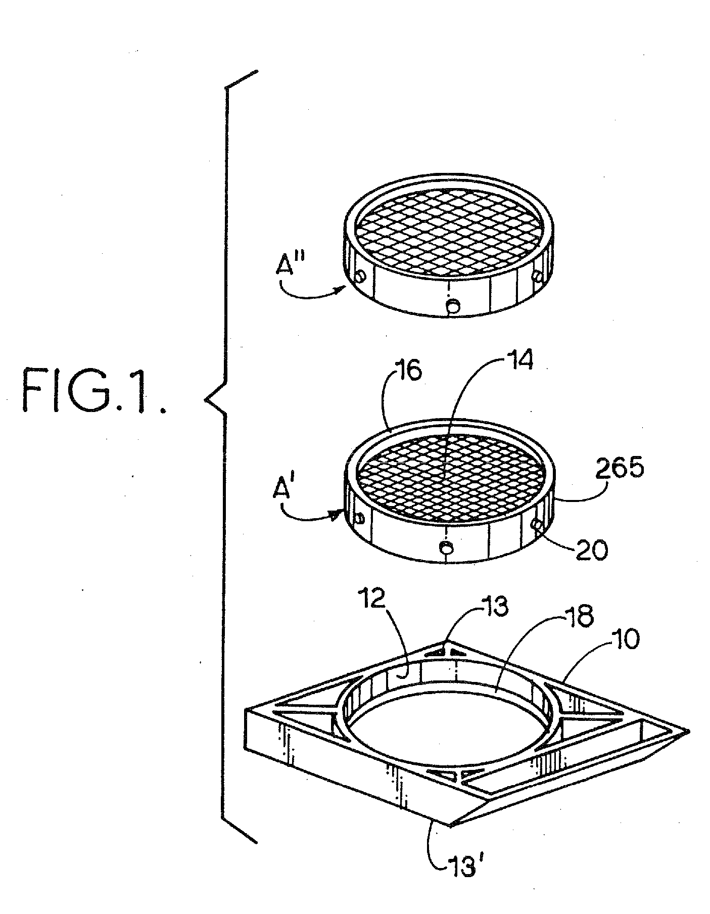
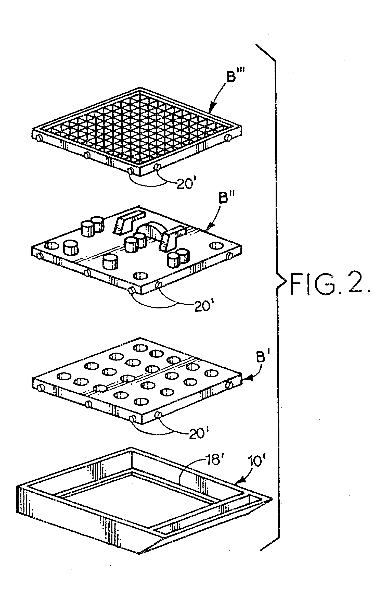
[0173] A platform includes a filter or stage assembled in a filter cassette frame or a stage cassette frame. FIGS. 1 and 2 show platform assemblies with interchangeable microtome sectionable tissue trapping filters, sectionable immobilizing stages or non-sectionable immobilizing stages and cassette frames.

Microtome Sectionable Tissue Support

[0174]FIG. 1 shows a cassette frame 10 with a cylindrical interior frame 12 which is designed to accept microtome sectionable tissue support, such as filters A′ and A″ each of which can be porous and forms a tissue support 14 surrounded by a collar 16. As discussed above, the term “filter” will be used for the tissue support because, in one form of the tissue support, fluid can pass through the tissue support while tissue samples are retained on the support in the manner of a filter. Tissue support 14 supports tissue samples during tissue processing, embedding and microtome and can include sectionable filters which c...


PUM


Abstract

A sectionable cassette for use in a process for harvesting and handling tissue samples for biopsy analysis is disclosed. In the procedure, a tissue biopsy sample is placed on a tissue trapping and supporting material that can withstand tissue preparation procedures and which can be cut with a microtome. The tissue is immobilized on the material and the material and the tissue are held in the cassette and are subjected to a process for replacing tissue fluids with wax, and then the tissue and the supporting material are sliced for mounting on slides using a microtome. Harvesting devices and containers using the filter material are disclosed. An automated process is also disclosed. One embodiment has the tissue trapping and supporting material porous and another embodiment includes a tissue supporting material that is not easily microtomed.

Description

CROSS REFERENCE TO RELATED APPLICATION [0001] This application is a divisional of application Ser. No. 09 / 890,177, filed Jul. 27, 2001 (pending), which is a 35 U.S.C. § 371 filing of PCT Application No. PCT / US98 / 20478, filed Oct. 5, 1998 (expired), which is a continuation-in-part of application Ser. No. 08 / 645,750, filed on May 14, 1996 (now U.S. Pat. No. 5,817,032), the disclosures of which are hereby fully incorporated by reference herein.TECHNICAL FIELD OF THE INVENTION [0002] The present invention relates to the general art of analysis of tissue samples, and to the particular field of obtaining, handling and processing tissue biopsy samples. Background of the Invention [0003] When disease is suspected in a living being, the physician must arrive at a specific diagnosis. Some disease processes, particularly tumors, require a histologic and / or cytologic diagnosis. While radiologic tools are useful in detecting the presence of a tumor, the cell type of the tumor can only be determi...

Claims


Application Information

Patent Timeline
IPC IPC(8): G01N1/00
CPCG01N1/286Y10T436/25G01N1/36
Inventor WILLIAMSON, WARREN P. IVWHITLATCH, STEPHENDINOVO, DOMINICALLEN, DOUGLASWARD, THOMAS
Owner BIOPATH AUTOMATION
Learn More