Method for detecting neoplastic disorders in a solubilized body sample

a solubilized body and neoplastic technology, applied in the field of solubilized body sample neoplastic disorders detection, can solve the problems of high inter- and intra-observer variance of experienced examiners, unsatisfactory false positive and false negative results of screening tests, and the inability to overcome the problem of preservation and preparation of samples by simply adding molecular markers, etc., to achieve effective and reliable kits and improve the morphological evaluation of cytology specimens

Active Publication Date: 2007-08-16
VENTANA MEDICAL SYST INC
View PDF40 Cites 26 Cited by
  • Summary
  • Abstract
  • Description
  • Claims
  • Application Information

AI Technical Summary

Benefits of technology

[0011] A method for detection of HPV nucleic acids from LBC samples is disclosed by Digene Corp. This method uses LBC samples as basis for the analysis. Detection of the HPV nucleic acids is performed after lysis of the cells contained in the LBC samples. In this method no normalization of the amount of the LBC sample to be employed in the biochemical non-cell based detection of the HPV nudeic acid, is performed with respect to information obtained from the cytological specimen prepared out of the same LBC sample. The method disclosed by Digene is therefore restricted to mere qualitative measurements. Any biochemical non-cell based quantitative or even semiquantitatve method needs information on the composition of the samples obtainable either from biochemical markers or from the microscopic or flow cytometric analysis of the sample. In the present invention the use of LBC samples for the assessment of diagnosis or for development of kits and in-vitro diagnostic devices enables for an accurate and comparable way to provide cytological information for the biochemical non-cell based testing. The employment of biochemical normalization with respect to markers indicative for the presence or absence of cells or cell types is omissible. The advantage of using LBC samples in this respect is that the cytologically cell based information is direct related to the homogeneous LBC specimen and thus provides valuable accurate information for use in the evaluation of the biochemical non-cell based test results.
[0012] A method for detection of molecular markers on the protein or nucleic acid level from solubilized specimens on the other hand is disclosed in various publications. However no link to the use of LBC samples as a source of the sample specimen in made in this respect. Generally LBC methods are applied in the art to enable for improved morphological evaluation of cytology specimens. The field of application of the LBC samples is therefore indicated only for cyto...

Problems solved by technology

Thus, for examination of cervical swabs and smears cytologists and pathologists have to be especially trained, and even experienced examiners have a high inter- and intra-observer variance in the assessment of a diagnosis based on cytological specimens.
As a result, the rate of false positive and false negative results in the screening tests remains unsatisfying high.
Yet the problem with the preservation and preparation of the samples may not be overcome by just additionally using molecular markers.
One further complication when performing cytological or histological examinations for screening purposes and especially when applying methods for the detection of molecular markers originates from strict precautions in preserving the samples from causing artefacts or improper results.
This is in part due to the instability...

Method used


Image

  • Method for detecting neoplastic disorders in a solubilized body sample

Examples


example 1

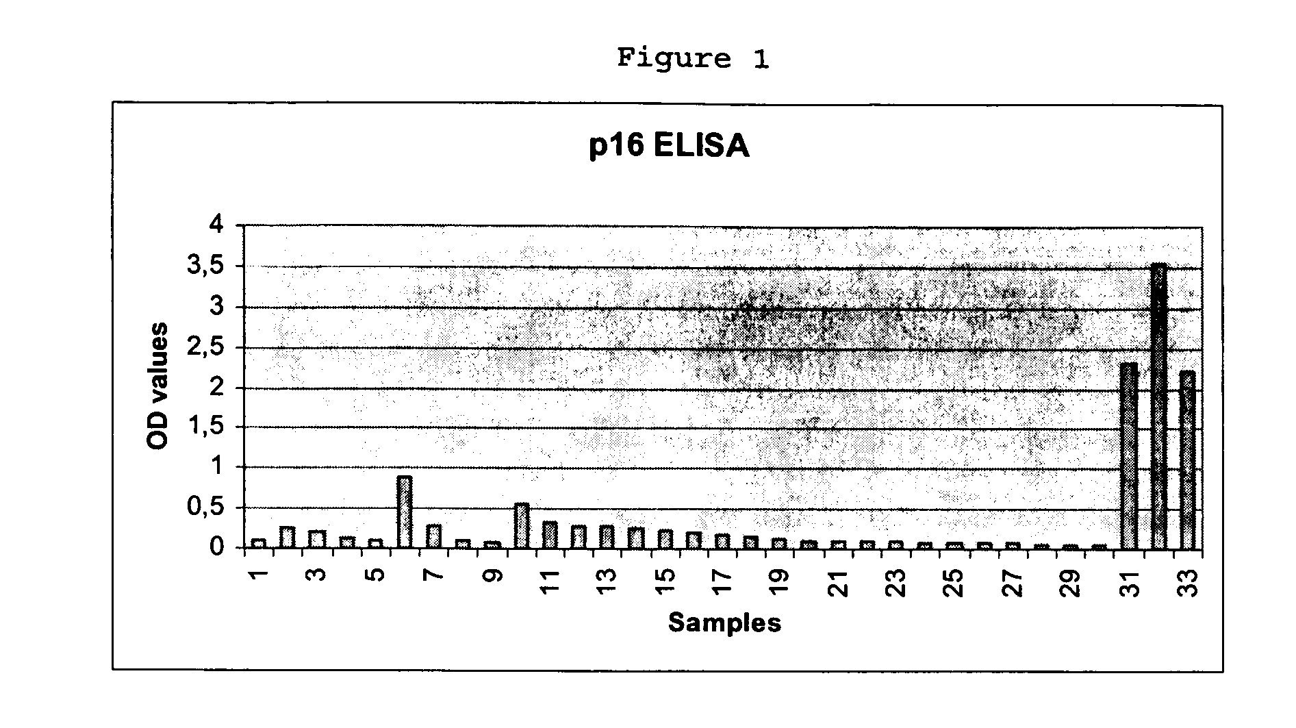
Detection of Cervical Intraepithelial Neoplasia in an ELISA Test Format

[0161] 33 cervical swabs provided in a lysis medium were subjected to ELISA based detection of overexpression of cyclin-dependent kinase inhibitor p16INK4a in solutions prepared from the cells contained in the swabs. The ELISA testing was performed as follows:

(A) Cell Lysis

[0162] Cervical swab brushes were given into 15 ml vessels, containing 2 ml of mtm lysis medium (2% Triton X-100, 0.4% SDS, 0.6 mM PMSF in PBS). Cervical cells present in the brush were lysed for at least 20 h. The lysates of the cervical swab samples were then transferred in 2 ml tubes and were centrifuged at 4° C. (15 min at 28.000×g (16.600 rpm Highspeed Centrifuge JEC Multi RF)); -Supernatant was transferred to a fresh tube. The Supernatant may be stored at −20° C.

(B) Performing the ELISA

[0163] Coating of ELISA-plates

[0164] Stock-solution of p16INK4a specific antibody clone mtm E6H4 was diluted in PBS to give ready-to-use coating so...

example 2

Detection of Cervical Intraepithelial Neoplasia in an Lateral Flow Test Format

[0194] Nine cervical swabs provided in PreservCyt (Cytyc Corporation, Boxborough, Mass.) solution have been subjected to conventional PAP testing and simultaneously to lateral flow based detection of overexpression of cyclin-dependent kinase inhibitor p16INK4a in solutions prepared from the cell suspensions obtained from the swabs. The lateral flow testing was performed as follows:

[0195] (A) Cell Lysis

[0196] 10 ml of the cell suspensions from the individual cervical swab samples provided as PreservCyt™ fixed materials were transferred to a 15 ml reaction vessel. The samples were centrifuged 15 min at ambient temperature at 1500×g (3000 rpm, Heraeus Varifuge, rotor 8074); supernatant was discarded, and remaining methanol allowed to evaporate (15 min at ambient temperature); the pellet was solubilized in 500 μl Lysisbuffer and transferred to a 1.5 ml reaction vessel. The solution was centrifuged at 4° C. ...

example 3

Detection of p16INK4a and p14ARF Transcripts by RT-PCR,

[0208] Cervical samples from 50 individuals were used for this analysis. For each individual two samples were obtained, one in Universal Collection Medium and one in PreservCyt™ solution. Both samples were obtained during the same examination session. For each of the individuals a diagnosis based on analysis of a cervical thin layer specimen prepared out of the PreservCyt™ solution was available. 20 of the samples included in the present study were chosen to be diagnosed as NILM, 20 samples were chosen to be LSIL and 10 samples were chosen to be HSIL. From all samples the level of transcripts of p16INK4a and of p14ARF have been determined on an mRNA level by RT-PCR according to the following protocol:

[0209] For performance of the analysis the cells were pelleted from the UCM and PreservCyt™ solutions by centrifugation. The pellets obtained were directly subjected to the RNA preparation procedure.

[0210] The pellet was diluted ...


PUM

PropertyMeasurementUnit
Electroluminescenceaaaaaaaaaa
Levelaaaaaaaaaa
Chemiluminescenceaaaaaaaaaa

Abstract

The present invention relates to a method for the early diagnosis of neoplastic disorders such as cancers as well as their precursor stages, particularly cancers of the respiratory tract, the urinary system, the reproductive tract, cancer associated with HPV infection or cancer of the anogenital tract, from solubilized body samples. The invention is also directed to test kits usable for this purpose as well as in-vitro diagnostic devices. The development of the kits and in-vitro diagnostic devices for the above purpose is also one aspect of the present invention.

Description

[0001] The present invention relates to a method for the early diagnosis of neoplastic disorders such as cancers as well as their precursor stages, particularly cancers of the respiratory tract, the urinary system, the reproductive tract, cancer associated with HPV infection or cancer of the anogenital tract, from solubilized body samples. BACKGROUND OF THE INVENTION [0002] Preventive programs have been offered for the most differing cancers since the middle of the fifties. For cervical cancer an established population wide screening program exists in various developed countries. However similar screening programs are applicable for other cancer entities and the respective precursor stages such as e.g. cancers of the urinary system, of the respiratory tract and other. In the following cervical cancer is used as an example to highlight the drawbacks of the present preventive scenario. However the facts are mutandis mutatis applicable to other preventive programs for any cancer entity...

Claims


Application Information

Patent Timeline
IPC IPC(8): C12Q1/68G01N33/574
CPCG01N33/57411Y10T436/25G01N33/57484
Inventor RIDDER, RUEDIGERREICHERT, ANJAVON KNEBEL DOEBERITZ, MAGNUSHERKERT, MATTHIASDUWE, ALEXANDERHIPFEL, RAINERMARTIN, PETER
Owner VENTANA MEDICAL SYST INC
Why Eureka
Try Eureka