Multi-leaf collimator based field size clipping for automatic adaptation to allowed image area

a collimator and field size technology, applied in the field of radiotherapy devices, can solve the problems of reducing the usability of the portal imaging system, damage or degrade,

Inactive Publication Date: 2007-08-23
SIEMENS MEDICAL SOLUTIONS USA INC
View PDF18 Cites 11 Cited by
  • Summary
  • Abstract
  • Description
  • Claims
  • Application Information

AI Technical Summary

Problems solved by technology

This active imaging area is typically surrounded by radiation sensitive electronics, which if exposed to ...

Method used


Image

  • Multi-leaf collimator based field size clipping for automatic adaptation to allowed image area
  • Multi-leaf collimator based field size clipping for automatic adaptation to allowed image area
  • Multi-leaf collimator based field size clipping for automatic adaptation to allowed image area

Examples


Embodiment Construction

[0021] Reference will now be made in detail to the presently preferred embodiments of the invention, examples of which are illustrated in the accompanying drawings.

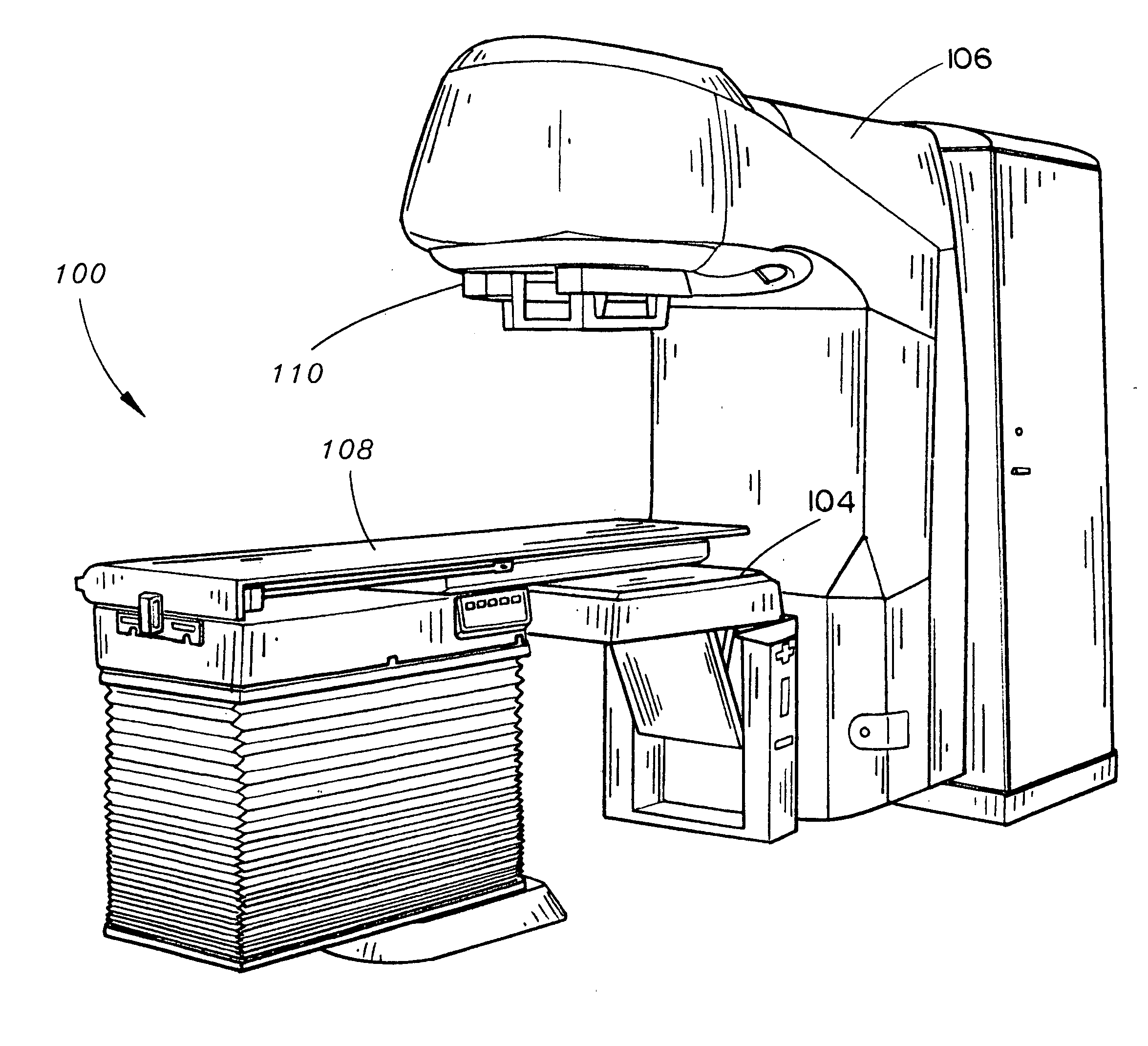
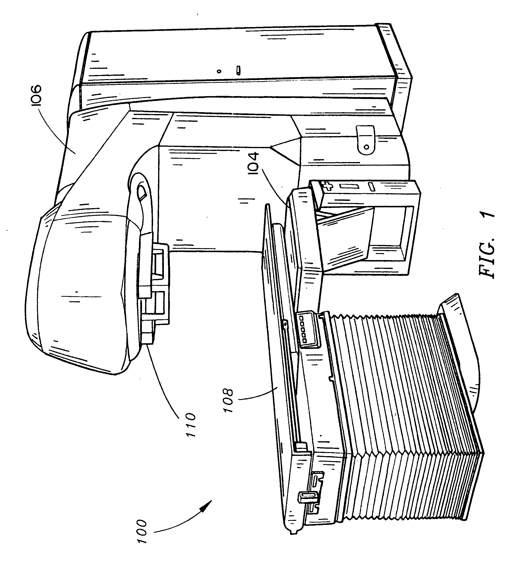
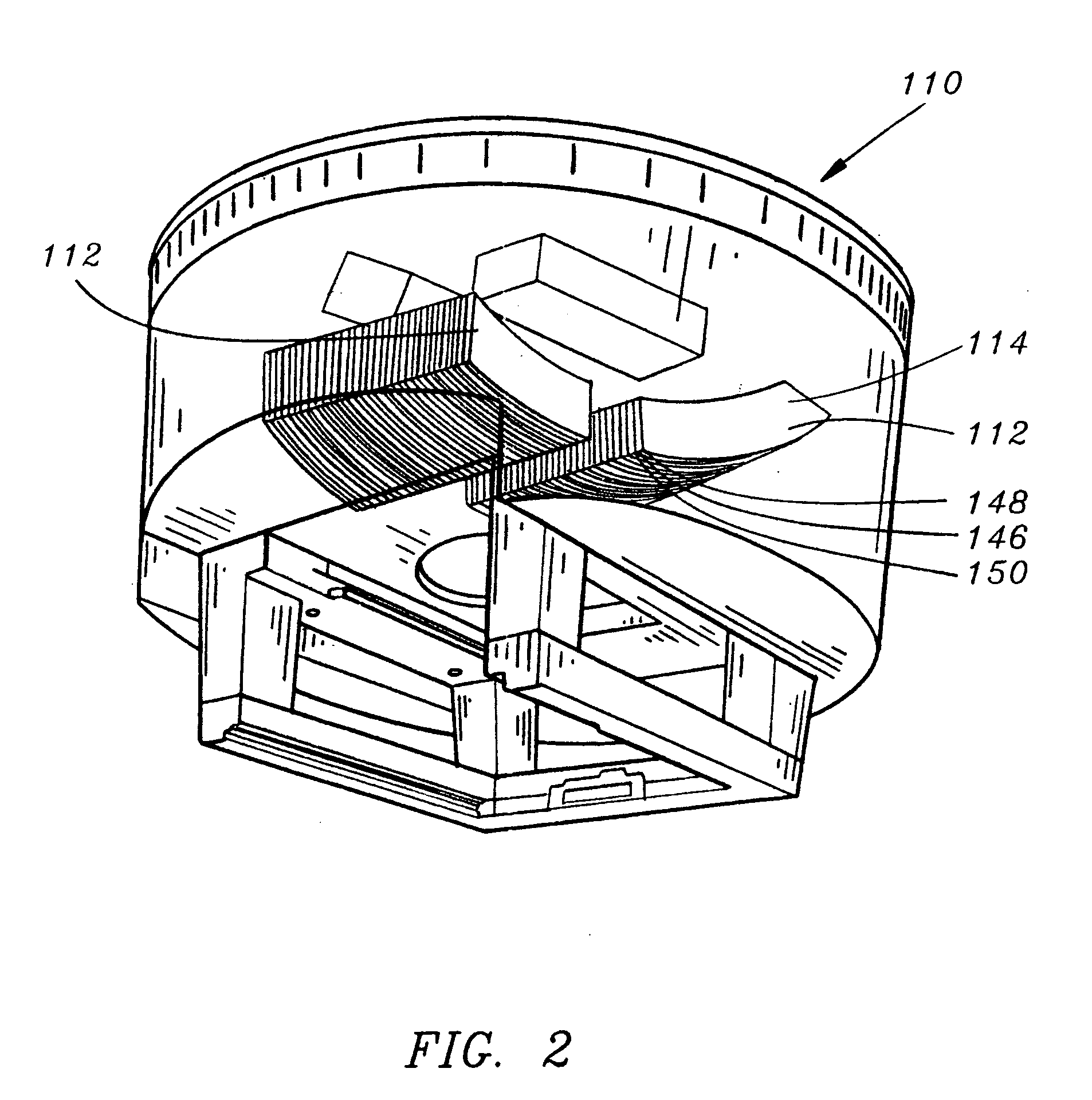
[0022] Referring generally to FIGS. 1 through 12, a system and method is described for capturing a portal image using a linear accelerator having a multi-leaf collimator and an imager, wherein the portal image field may be automatically clipped to account for the allowed image area of the imager using the multi-leaf collimator in accordance with an exemplary embodiment of the present invention. In this manner, the present invention provides for automatic optimization of a selected portal image field size to accommodate the dimensions of the imager. It will be appreciated that clipping of the portal image field in accordance with the present invention has no impact on the clinical usage of the linear accelerator since no clinical information is lost due to clipping of the portal image field. The projection of the clipped ...


PUM


Abstract

A system and method for capturing a portal image using a linear accelerator having a multi-leaf collimator (MLC) and an imager allows the portal image field to be automatically clipped using the multi-leaf collimator so that the projection of the portal image field onto the imager falls within the allowed image area of the imager. In this manner, portal images may be obtained in instances where the projection of the portal image field onto the flat-panel imager is larger than an allowed image area of the imager, or where the projection of the portal image field onto the imager is not centered and would extend beyond the allowed image area, so that radiation sensitive electronics outside of the allowed image area are protected from undesirable exposure to radiation.

Description

BACKGROUND OF THE INVENTION [0001] The present invention relates generally to the field of radiation therapy devices such as medical linear accelerators equipped with portal imaging systems, or the like, typically used for providing Image Guided Radiation Therapy (IGRT), and more particularly to a system and method for providing automatic field size clipping to the allowed image area of the imager of the portal imaging system using the multi-leaf collimator of the linear accelerator. [0002] IGRT uses patient positioning devices and radiation imaging technologies such as portal imaging to target and treat cancerous tumors more precisely. Prior to the use of IGRT, radiation oncologists contended with variations in patient positioning, including variations caused by a patient's respiratory motion. Inevitably, a margin of healthy tissue around a treatment site was treated with radiation. However, the use of IGRT allows a radiation oncologist to determine the exact positioning of a treat...

Claims


Application Information

Patent Timeline
IPC IPC(8): G21K1/04A61N5/10
CPCA61N5/1042A61N5/1065G21K1/046G21K1/04A61N2005/1054
Inventor MANTHEY, DIETERPOND, DAVESTAHL, JOHANNESAHUMADA, RICARDO
Owner SIEMENS MEDICAL SOLUTIONS USA INC
Why Eureka
Try Eureka