Medical devices with enhanced ultrasonic visibilty

a technology of enhanced ultrasonic visibilty and medical devices, which is applied in the field of medical devices, can solve the problems that the ultrasonic imaging system may no longer be able to detect and display

Inactive Publication Date: 2007-08-23
ARTENGA
View PDF6 Cites 896 Cited by
  • Summary
  • Abstract
  • Description
  • Claims
  • Application Information

AI Technical Summary

Problems solved by technology

As a needle is inserted to a depth within a patient, an ultrasound imaging system may no longer be able to detect and display it.

Method used


Image

  • Medical devices with enhanced ultrasonic visibilty
  • Medical devices with enhanced ultrasonic visibilty
  • Medical devices with enhanced ultrasonic visibilty

Examples


Embodiment Construction

[0068] Reference will now be made in detail to various suitable embodiments of the invention as illustrated in the accompanying drawings. It will be understood that this description is exemplary and is to assist in understanding the invention and the principles of operation.

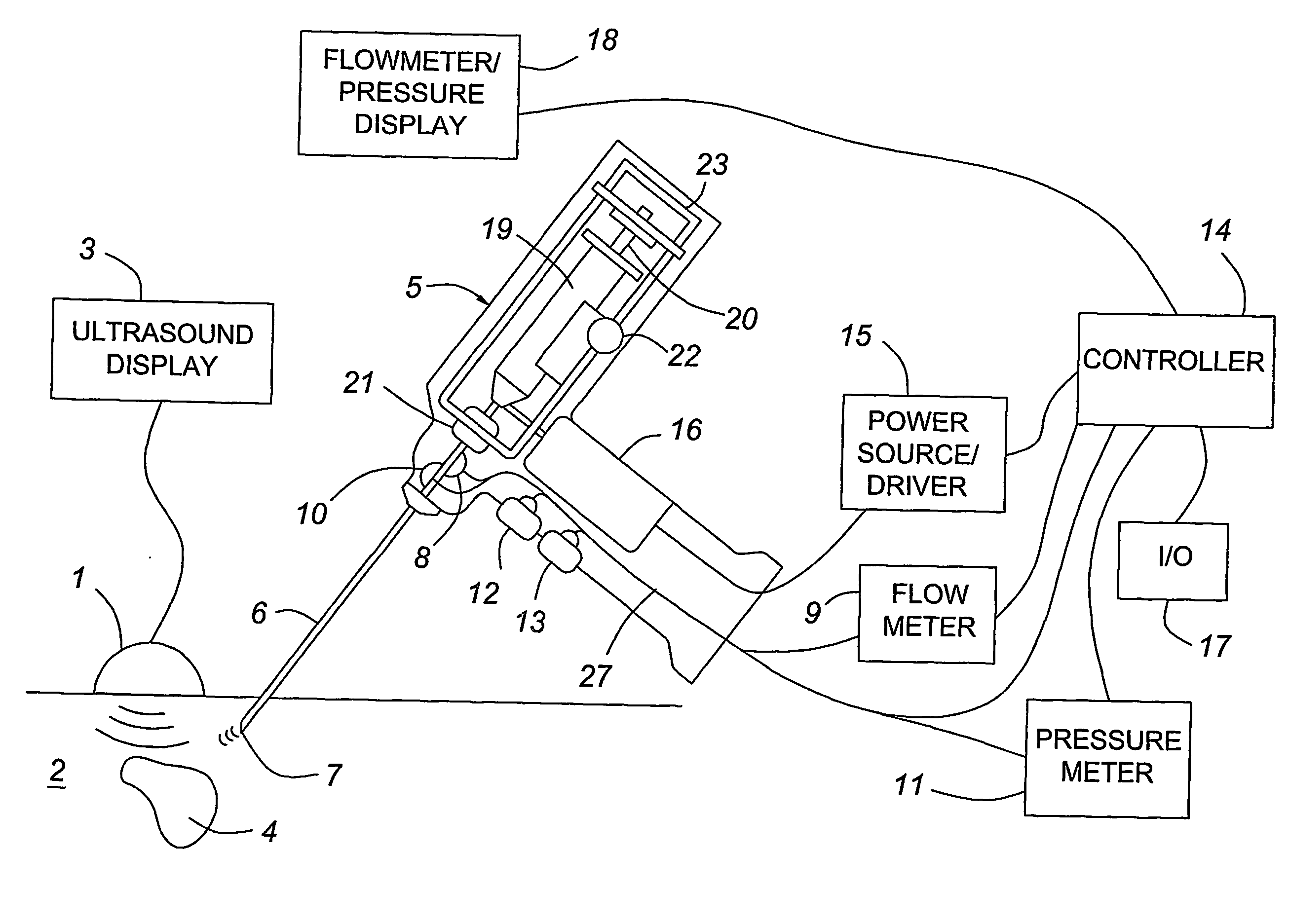
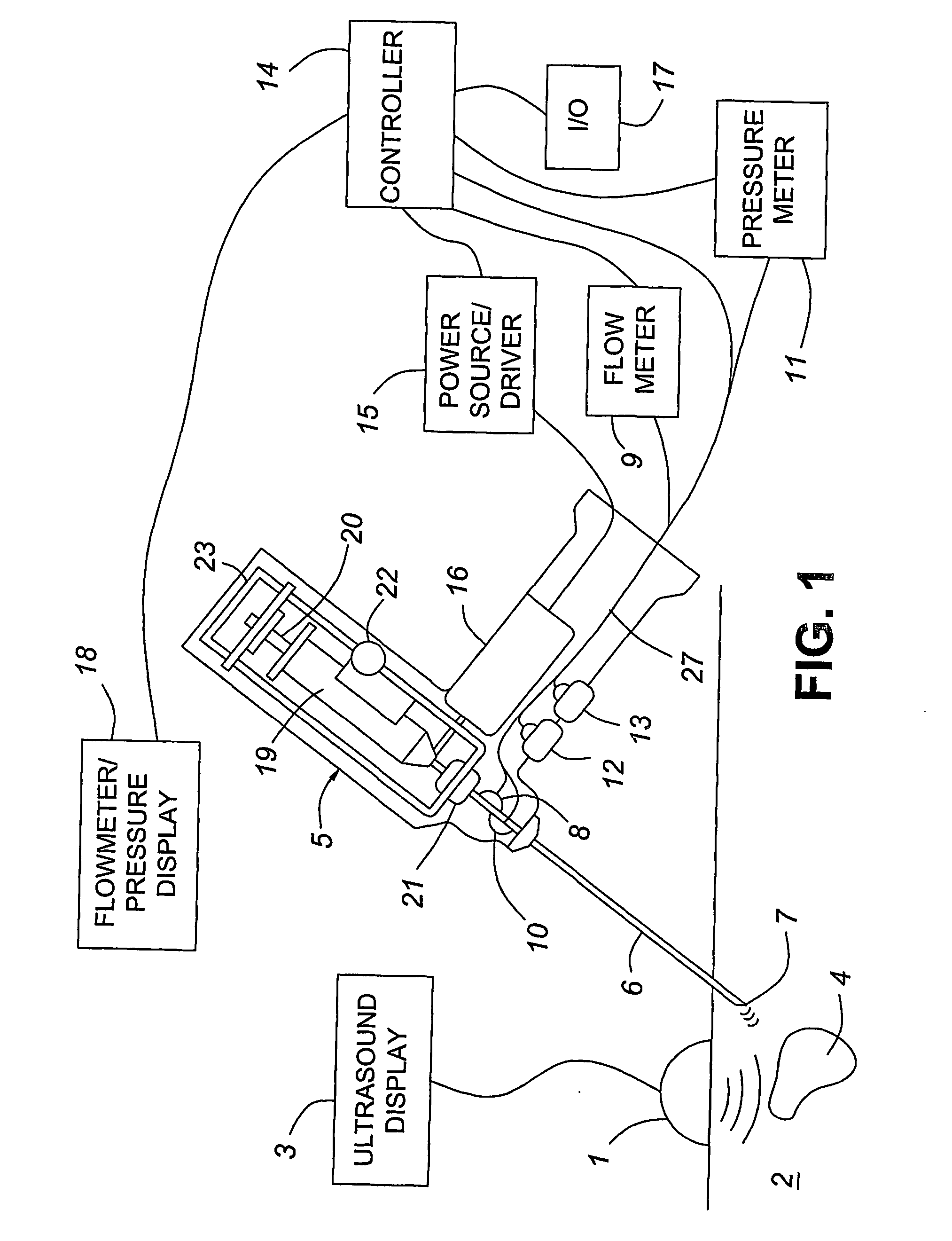
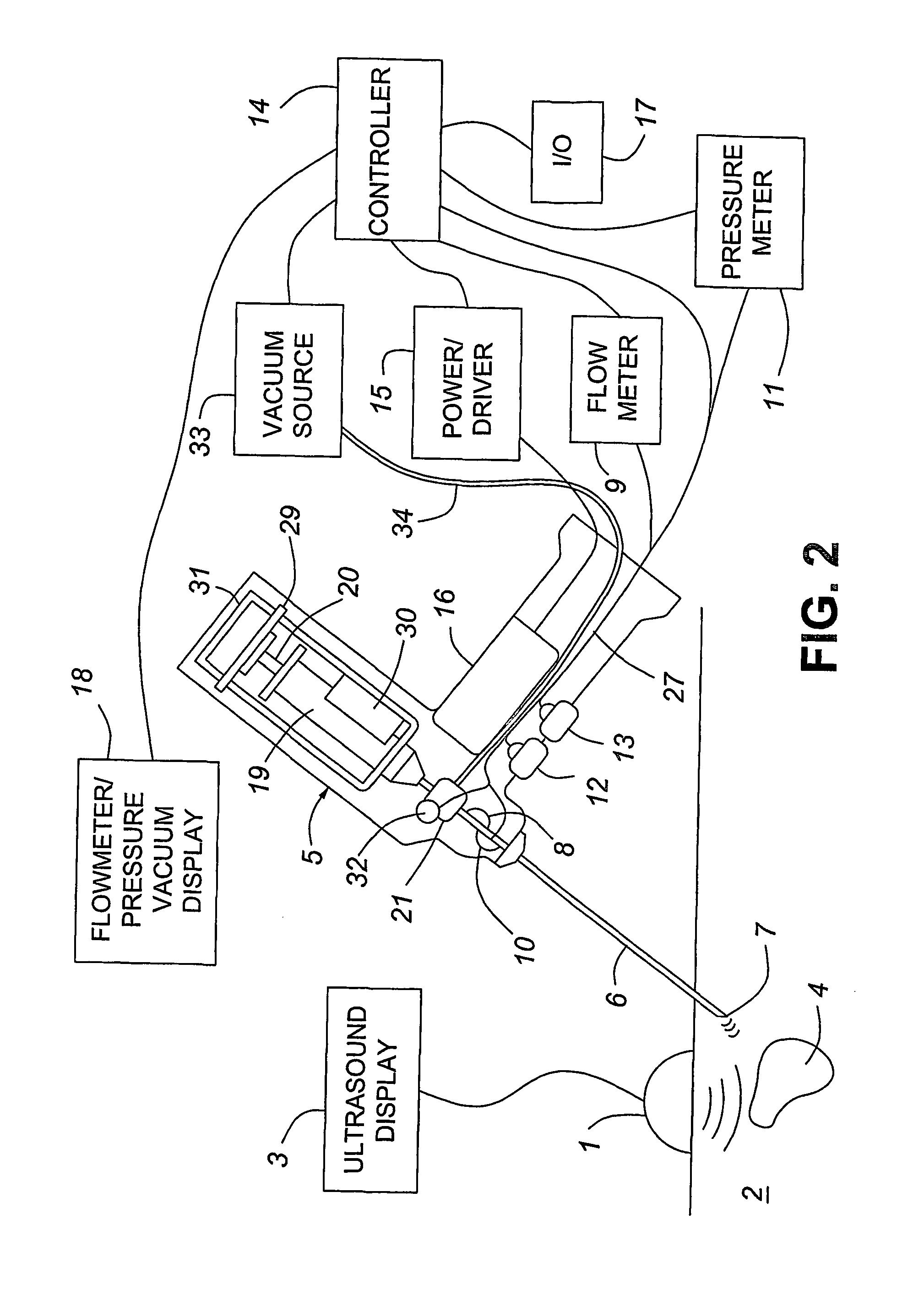
[0069] Devices of the present invention includes means for providing an echogenic fluid from a needle, and analysis thereof, to enhance the ultrasonic visibility of the needle tip. The device may be comprised of a handheld assembly or of a system, comprised of a handheld assembly connected to other components such as fluid vessels, power sources, and meters.

[0070] The term needle is intended to include any hollow, slender instrument that may be manipulated to puncture or be inserted or otherwise probe tissues, organs, cavities, or the like. The needle may be used to introduce material into or remove material from a patient or to perform other therapeutic or diagnostic functions. The term needle is intended to i...


PUM


Abstract

A medical device having enhanced ultrasonic visibility is provided. The device permits localized drug delivery, probe positioning, fluid drainage, biopsy, or ultrasound pulse delivery, through the real-time ultrasound monitoring of the needle tip position within a patient. The device permits controlled dispersion of a drug into solid tissue, the lodging of particles into solid tissue, and drug delivery into specific blood vessels. As a needle is inserted, a fluid that contrasts echogenically with the organ environment is injected into the patient. The fluid travels a brief distance before being slowed and stopped by the patient's tissue and this fluid flow will be detectable by ultrasound. The needle position during insertion will be monitored using ultrasound until it is at the desired point of action. A therapeutic drug is then delivered or a probe inserted

Description

FIELD OF THE INVENTION [0001] The invention generally pertains to medical devices employing ultrasonic guidance of positioning. Such devices can be used, for example, to probe, inject therapeutic agents, drain bodily fluids, perform biopsy or to provide diagnostic imaging agents. Such devices can be used to enhance the dispersion of a therapeutic agent into solid tissue as well as to deliver a therapeutic agent into specific blood vessels. Such devices can be used to precisely position a probe within a patient in order to permit solid tumor ablation through heat, freezing, or brachytherapy. BACKGROUND TO THE INVENTION [0002] Medical Rationale [0003] Accurate, real-time knowledge of a needle tip location is an obvious requirement of a biopsy procedure. It is also desired in order to deliver drugs to a specific target site as well as to avoid puncture damage to other tissue. [0004] Biotherapeutics, which are expected to comprise more than half of the new drugs developed in the next tw...

Claims


Application Information

Patent Timeline
Patent Type & Authority Applications(United States)
IPC IPC(8): A61N1/30A61B8/00A61B10/00A61B17/22A61B17/34A61B19/00A61K9/127A61K45/00A61K49/00A61M1/00A61M5/00A61M5/145A61M5/32A61M27/00
CPCA61B8/0833A61B8/0841A61B8/481A61B17/3468A61M2205/3331A61B2017/22088A61B2017/22089A61B2019/5425A61M5/1456A61B2017/22008A61B2090/3925
Inventor KEENAN, JAMES
Owner ARTENGA
Why Eureka
Try Eureka