Synthetic microfluidic microvasculature network

a microfluidic microvasculature and synthetic technology, applied in the field of synthetic microfluidic microvasculature network, can solve the problems of inability to predict, inability to provide realistic sizes and geometries corresponding, and inability to understand the complex interplay between flow, cells and particles, etc., to eliminate concerns of cross-contamination and optimize drug delivery

Active Publication Date: 2007-10-04
SYNVIVO INC
View PDF1 Cites 16 Cited by
  • Summary
  • Abstract
  • Description
  • Claims
  • Application Information

AI Technical Summary

Benefits of technology

[0017] The present invention provides apparatus and methods that can be used to study fluid flow and particle adhesion in physiological vessels including arterioles, capillaries, venules, and microvascular networks comprising any combination of the three. The same apparatus and methods can also be used to optimize drug delivery in the microvasculature. Many drugs are available in microencapsulated, liposomal / lipisomal, and other micro and nanoscale particle forms. The adherence and uptake of these particles in the microvasculature depends on specific and non-specific interactions between the surfaces of drug delivery particles and endothelial cells that line the walls of the microvasculature. Adhesion also depends on fluid dynamics parameters such as flow velocities and shear forces which, in turn, depend on vascular network geometries. The present invention provides microfluidic chips comprising synthetic microvascular networks (SMNs) with flow channels that possess key geometric and topological features that cause them to display the same types of fluid flow patterns and particle adhesion patterns as are found in physiological microvascular networks. In addition, the SMNs require quantities of reagents that are reduced by orders of magnitude compared with currently used techniques. Known microfabrication techniques also allow for development of plastic, disposable chips eliminating concerns of cross-contamination.

Problems solved by technology

This complex interplay between flow, cells, and particles is still poorly understood and it is not possible to predict, for example, adhesion patterns and numbers of adhered particles in the microvasculature based on current in-vitro flow cell technologies.
They do not provide realistic sizes and geometries corresponding to complex combinations of thoroughfare channels, bifurcations, junctions, convolutions, and / or variable diameters found in in-vivo microcirculation.
Existing in-vitro flow chambers, however, having idealized rectangular or circular duct geometries cannot be modified to reflect changes that occur with disease.
However, both studied idealized (straight) microchannels that did not account for either the interconnectedness or variations found in in-vivo microvascular networks.
All of the numerical studies mentioned above are based upon simple, idealized flow chambers with regular geometries and have not examined flow profiles and shear stresses in realistic in-vivo microvasculature scenarios with relevance to cell adhesion.

Method used


Image

  • Synthetic microfluidic microvasculature network
  • Synthetic microfluidic microvasculature network
  • Synthetic microfluidic microvasculature network

Examples


examples

CFD Simulation of Particle Transport and Adhesion in a SMN and Experimental Validation

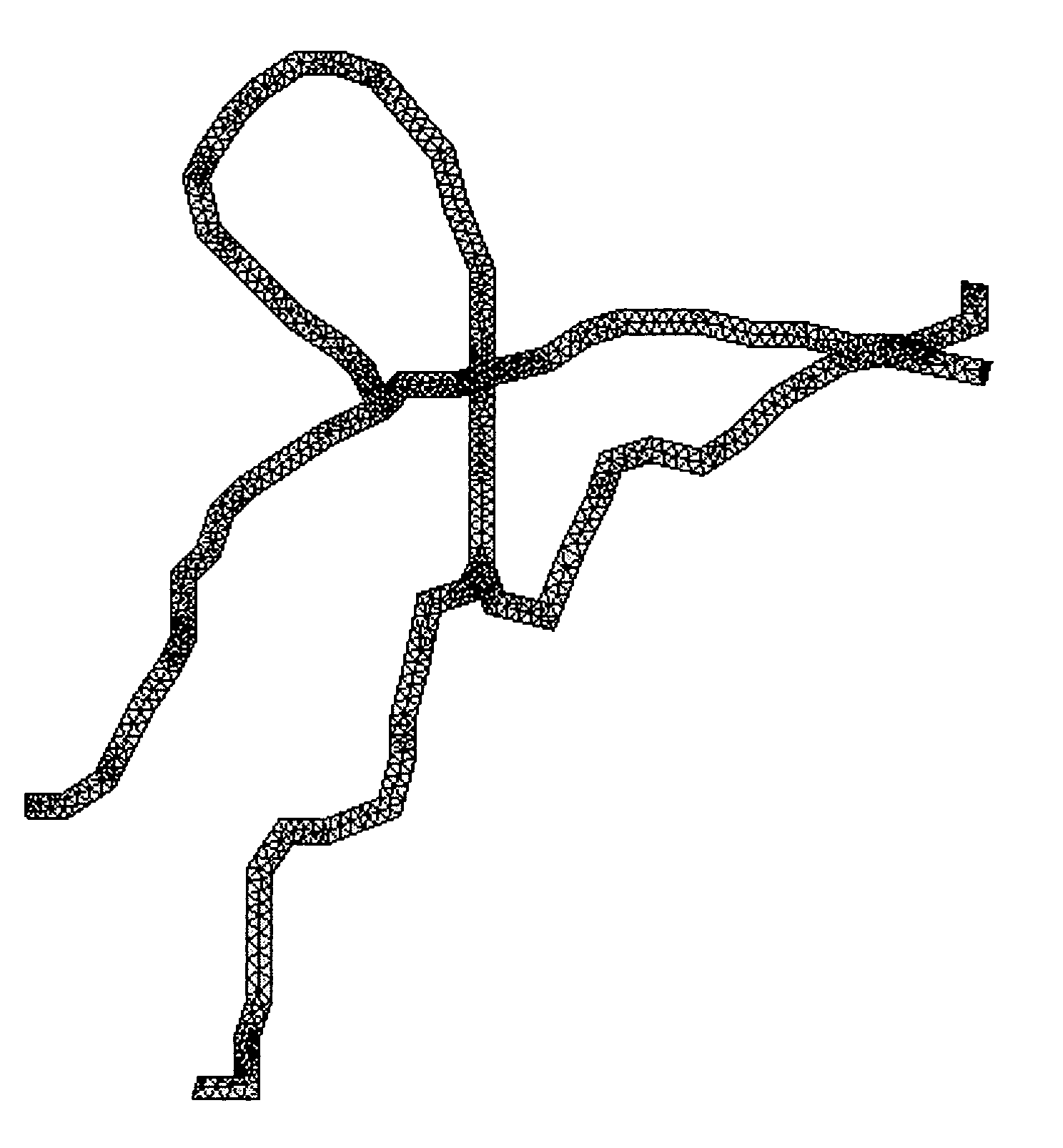
[0083] Particle transport at two junctions in the microvascular network shown in FIG. 12 is presented in FIG. 13 and FIG. 14. The direction of particle transport at the junction is indicated by arrows. Experimentally measured and numerically predicted values of particle flux at the two bifurcations shown in FIG. 13 and FIG. 14 are presented in FIG. 15. Fluid and particle flows are compared for both of the junctions in FIG. 16. The flow and particle ratios were defined with respect to the conditions in the arm labeled “In.” While the particle split is nearly identical to the flow split at Junction 2, the flow / particle split is significantly different at Junction 1. These effects are representative of the complexities of the in-vivo environment and are clearly not realizable in existing parallel flow chambers.

[0084] Particle adhesion in the SMN studies compared very well with simulated results. FIG...


PUM

PropertyMeasurementUnit
diametersaaaaaaaaaa
diametersaaaaaaaaaa
diametersaaaaaaaaaa

Abstract

The present invention is a synthetic microfluidic microvasculature network and associated methods. The synthetic microfluidic microvasculature network mimics the structure, fluid flow characteristics, and physiological behavior of physiological microvasculature networks. Computational methods for simulating flow and particle adherence in synthetic and physiological microvascular systems and methods for determining parameters influencing particle adhesion and drug delivery are also described. The invention has many uses including the optimization of drug delivery and microvascular treatments and in describing disease mechanisms that affect the microvasculature such as inflammation, diabetes and hypertension.

Description

STATEMENT REGARDING FEDERALLY SPONSORED RESEARCH OR DEVELOPMENT [0001] The U.S. Government may have certain rights in this invention pursuant to SBIR Contract Number 1R43HL076034-01 A1 awarded by the National Institutes of Health.CROSS-REFERENCE TO RELATED APPLICATIONS [0002] Not Applicable INCORPORATED-BY-REFERENCE OF MATERIAL ON A CD [0003] Not Applicable BACKGROUND OF THE INVENTION [0004] 1. Field of the Invention [0005] The present invention is in the field of microfluidics and physiology. More specifically, the invention relates to microfluidic systems that mimic the structure, fluid flow characteristics, and physiological behavior of small physiological vessels such as those found in the microvasculature. The invention has many uses including the optimization of drug delivery and microvascular treatments and in describing disease mechanisms that affect the microvasculature such as inflammation, diabetes and hypertension. The invention is not limited to vessels of the circulato...

Claims


Application Information

Patent Timeline
Patent Type & Authority Applications(United States)
IPC IPC(8): C12Q1/00G06F19/00C12M3/00
CPCG01N33/54346
Inventor PRABHAKARPANDIAN, BALABHASKARSUNDARAM, SHIVSHANKARPANT, KAPIL
Owner SYNVIVO INC
Why Eureka
Try Eureka