High resolution chromosomal mapping

a chromosomal mapping and high-resolution technology, applied in the field of mapping and structural analysis of chromosomes and chromosomal rearrangements, can solve the problems of limited spatial resolution, low resolution of chromosome boundaries, and inability to accurately and reproducibly determine the boundaries of genomic structures

Active Publication Date: 2007-10-11
AGILENT TECH INC
View PDF38 Cites 62 Cited by
  • Summary
  • Abstract
  • Description
  • Claims
  • Application Information

AI Technical Summary

Problems solved by technology

However these technologies are generally low resolution, and are often limited to the spatial resolution of the light microscope, e.g., several megabases.
Furthermore accurate and reproducible determination of the boundaries of genomic structures is limited by the relatively low resolution of these current techniques.

Method used


Image

  • High resolution chromosomal mapping
  • High resolution chromosomal mapping
  • High resolution chromosomal mapping

Examples


example 1

[0135] This example illustrates one method of mapping the boundaries of a genomic structure, according to one embodiment of the invention.

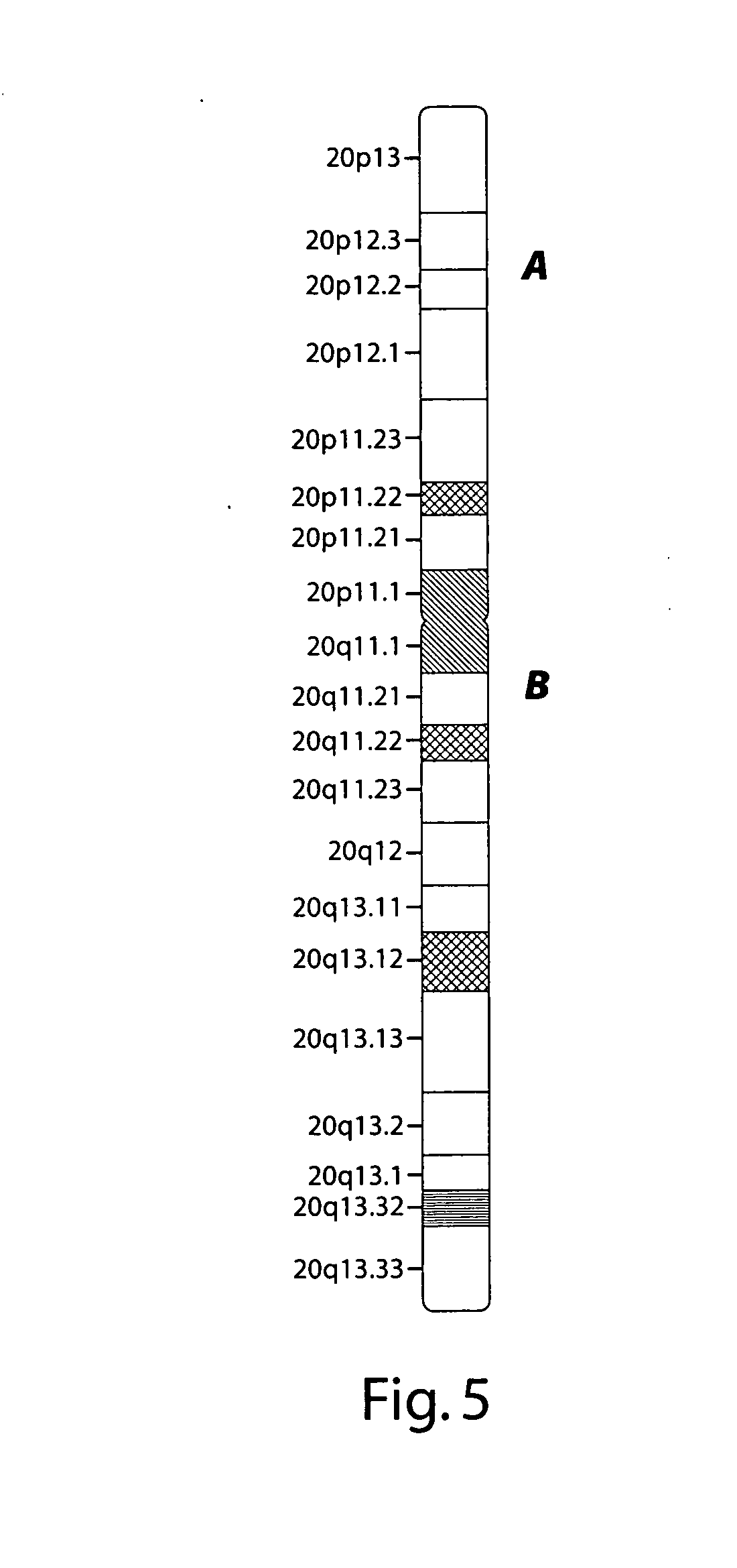
[0136] Chromosome 20 has 20 cytogenetic G-bands (20p13-20q13.33), as shown schematically in FIG. 5. A series of design probes of pools of long oligonucleotides were prepared. For the “p” arm, each pool included unique genomic sequences within a cytoband. The 5′ and 3′ ends of cytoband-specific pool precisely defined by DNA sequence.

[0137] For the “q” arm, each pool included unique genomic sequences of <100 kb within a single G band (e.g. 20q12). The 5′ and 3′ ends of each sub-cytoband pool were precisely defined by DNA sequence.

[0138] The sequences within individual cytobands can be precisely synthesized as individual pools of long oligonucleotides. These can also be used to provide high resolution mapping of any genomic structures within these regions.

[0139] It will be appreciated that throughout the present application, that words such as “c...


PUM

PropertyMeasurementUnit
temperatureaaaaaaaaaa
pHaaaaaaaaaa

Abstract

The present invention generally relates to spatial and structural genomic analysis compositions, which can be used for the mapping of chromosomes and structural analyses of chromosomal rearrangements, including the entire chromosome, as well as specific portions or regions of interest of the chromosomes. In some embodiments, multiple portions of the genome can be distinguished, for instance, using a first detection entity and a second detection entity different from the first detection entity. The detection entities may be immobilized relative to oligonucleotides, which may be selected to bind to different locations within the chromosome. For instance, the oligonucleotides may be at least substantially complementary to the chromosome, e.g., substantially complementary to a specific location of the chromosome.

Description

BACKGROUND [0001] Cytogenetics is typically used for identifying structures such as balanced translocations, marker chromosomes, and other genomic structures present in a sample of interest. Examples of cytogenetics techniques include Giemsa (G) banding, M banding, or spectral karyotyping (SKY). However these technologies are generally low resolution, and are often limited to the spatial resolution of the light microscope, e.g., several megabases. In addition techniques, such as M-banding require manual microdissection of the chromosomes of interest, e.g., under a light or fluorescence microscope. Therefore, structures of interest such as duplications, inversions, or translocations need to be at least several megabases (Mb) in length. Furthermore accurate and reproducible determination of the boundaries of genomic structures is limited by the relatively low resolution of these current techniques. Accordingly, there is a need to improve the spatial resolution of mapping and character...

Claims


Application Information

Patent Timeline
Patent Type & Authority Applications(United States)
IPC IPC(8): C12Q1/68C12P19/34C12M3/00
CPCC12Q1/6841
Inventor BARRETT, MICHAEL THOMASCAREN, MICHAEL P.
Owner AGILENT TECH INC
Why Eureka
Try Eureka