Transfer of treatment planning information using standard image transfer protocols

US20070238962A1Inactive Publication Date: 2007-10-11BRAINLAB

Image

  • Transfer of treatment planning information using standard image transfer protocols
  • Transfer of treatment planning information using standard image transfer protocols
  • Transfer of treatment planning information using standard image transfer protocols

Examples


Embodiment Construction

[0021]The embodiments described below relate generally to diagnostic medical images. Although any type of medical image can be used, these embodiments will be illustrated in conjunction with CT images. As noted in more detail below, other types of medical images can be used, and the following claims should not be limited to CT images unless explicitly recited therein.

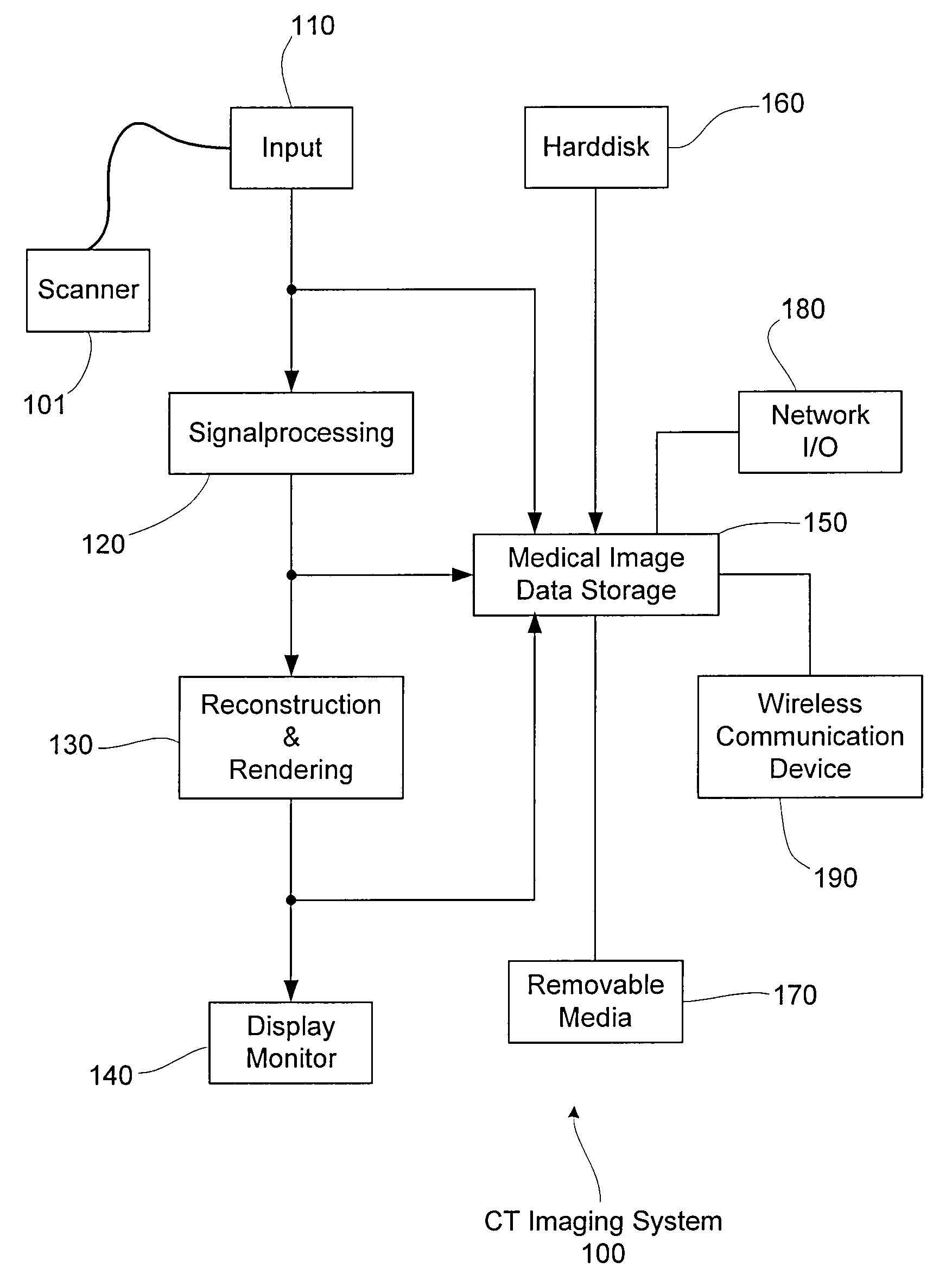
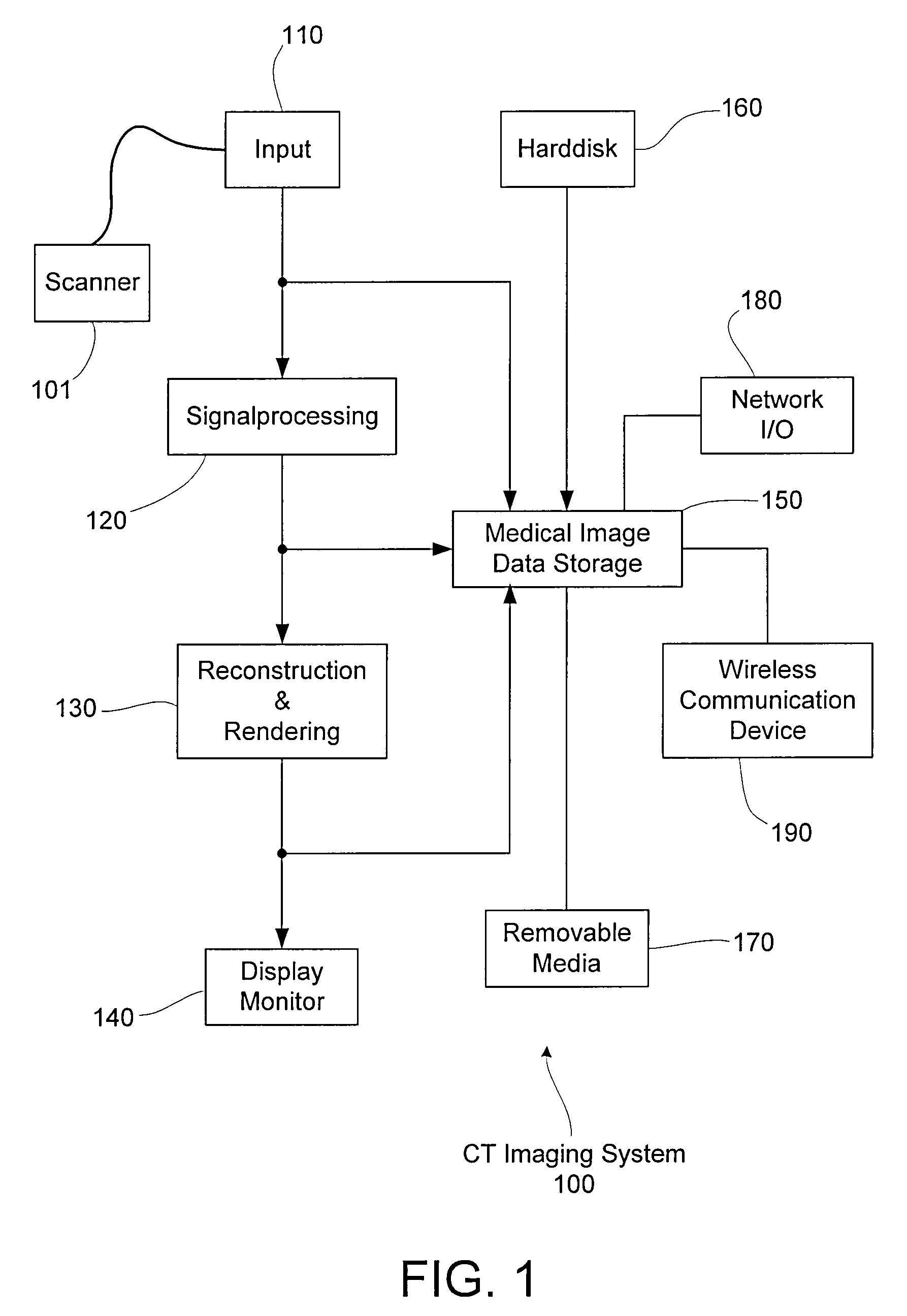
[0022]FIG. 1 is a block diagram of an exemplary CT imaging system 100. The CT imaging system 100 includes a CT scan data input port 110 connected to a CT scanner 101, a signal processing section 120, a reconstruction and rendering section 130, and a display monitor 140. The CT system 100 also includes a medical image data storage section 150, which can capture and / or store image data at one or more locations along the image path, a hard disk 160, removable media 170 (e.g., a CD, a DVD, etc.), a network I / O port 180, and a wireless communication device 190.

[0023]During a CT examination, the CT scanner 101 produces image ...


PUM


Abstract

A system and method for creating, generating, saving and / or exporting medical data is provided. The medical data includes image data, and the medical data conforms to an external standard, wherein additional data, not part of the external standard, is added to the image data. The additional data may be added by rendering the additional data into the image data to create new data including at least part of the original image data and at least a part of the additional data, wherein the new data conforms to the external standard.

Description

RELATED APPLICATION DATA[0001]This application claims priority of U.S. Provisional Application No. 60 / 745,031 filed on Apr. 18, 2006, which is incorporated herein by reference in its entirety.FIELD OF THE INVENTION[0002]The present invention relates to a method and a device for generating, exporting and / or saving data, such as medical image data. More particularly, the invention relates to including non-standard data (e.g., additional image information or data not part of the standard) into a data structure that conforms to a particular standard, such as DICOM.BACKGROUND OF THE INVENTION[0003]Since medical imaging equipment typically interoperates with other medical devices, it is common for medical scanners (e.g., CT, MR, PET, or SPECT scanners), other medical imaging devices, displays and / or software to be interoperable so as to exchange data based on a common standard or protocol. An example of such a protocol is the DICOM (digital imaging and communication in medicine) standard ...

Claims


Application Information

Patent Timeline
11 Oct 2007
Publication
US20070238962A1
IPC
A61B5/05
CPC
A61B6/032; G06F19/321; A61B6/563; A61B6/037; G16H30/20; G16H30/40
Inventors
HARTLEP, ANDREAS; FRIELINGHAUS, NITS