
Determination of a Measure of a Glycation End-Product or Disease State Using Tissue Fluorescence

a tissue state and fluorescence technology, applied in the field of tissue state determination, can solve the problems of poor sensitivity of fpg, 40-60% of late diagnosis, inadequate screening methods for type 2 diabetes and pre-diabetes, etc., and achieve the effect of reducing measurement time, increasing the overall signal to noise ratio, and improving detection accuracy

Inactive Publication Date: 2007-11-29
VERALIGHT INC
View PDF50 Cites 29 Cited by
  • Summary
  • Abstract
  • Description
  • Claims
  • Application Information

AI Technical Summary

Benefits of technology

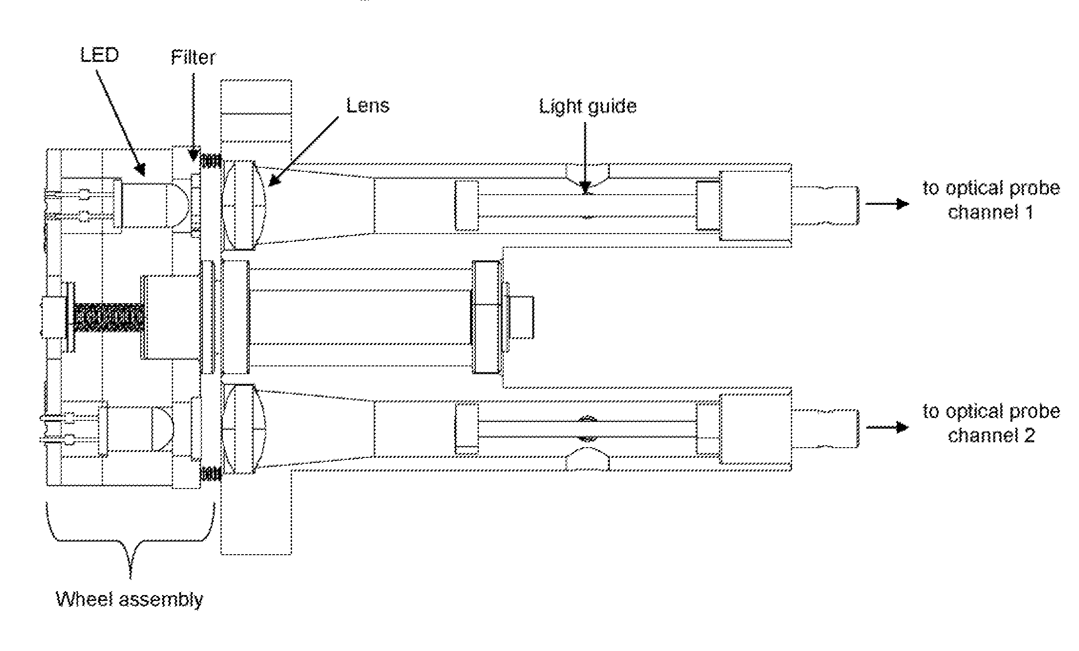
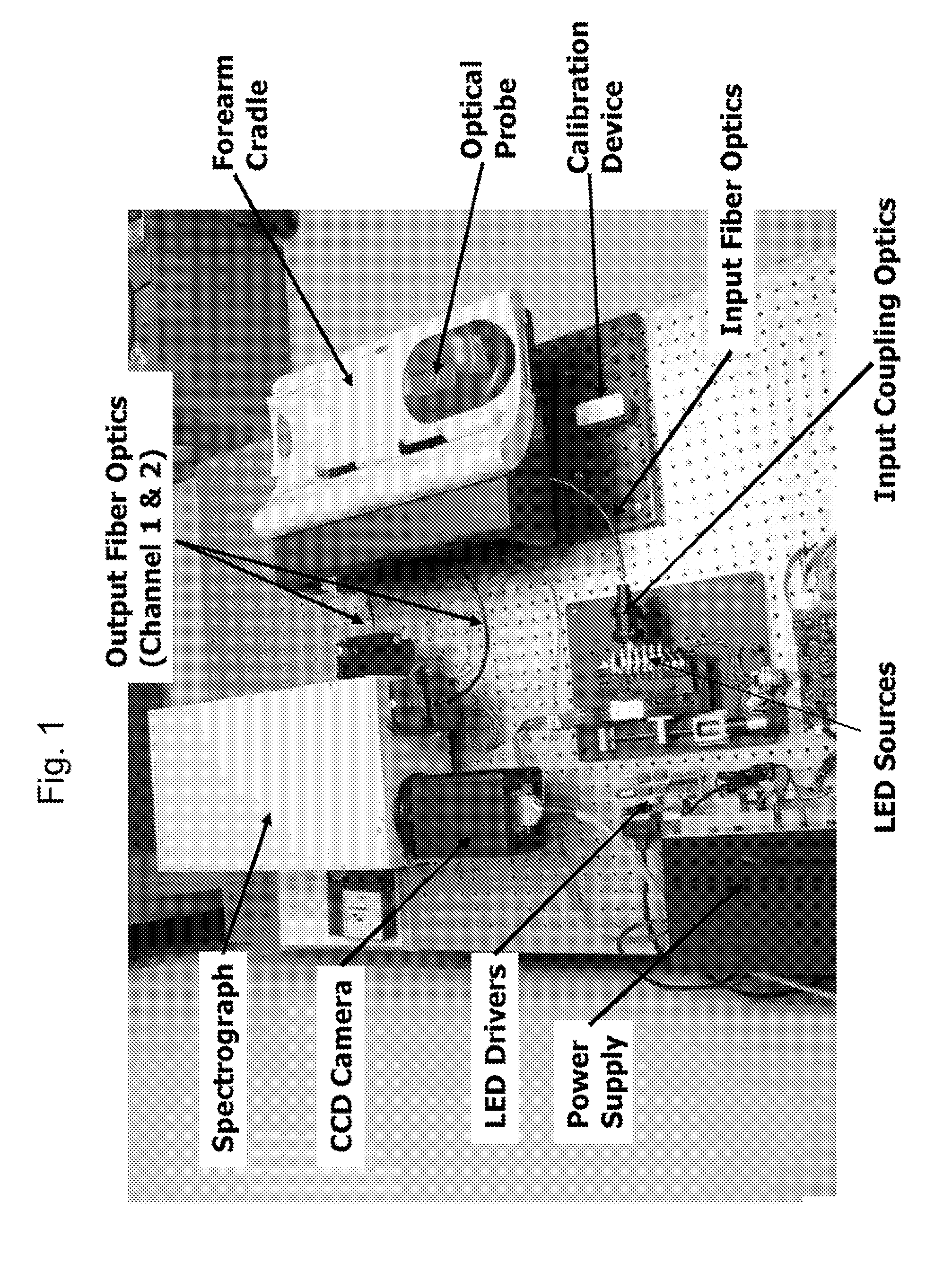
[0008] A noninvasive method and apparatus for detecting disease in an individual using fluorescence spectroscopy and multivariate analysis has been previously disclosed in U.S. Pat. No. 7,139,598, incorporated herein by reference. Continued development of this method and apparatus has resulted in significant instrument and algorithm improvements that yield increased accuracy for noninvasively detecting disease, especially type 2 diabetes and pre-diabetes. The instrument improvements provide higher overall signal to noise ratio, reduced measurement time, better reliability, lower cost and reduced size compared to instruments disclosed in the art. The algorithmic improvements improve overall accuracy by more effective extraction of the information needed for accurate noninvasive detection of disease using fluorescence spectroscopy. These instrument and algorithm improvements are described herein, and have been tested in a large clinical study also described herein.

Problems solved by technology

However, current screening methods for type 2 diabetes and pre-diabetes are inadequate due to their inconvenience and inaccuracy.
FPG also suffers from poor sensitivity (40-60%) contributing to late diagnoses.

Method used


Image

  • Determination of a Measure of a Glycation End-Product or Disease State Using Tissue Fluorescence
  • Determination of a Measure of a Glycation End-Product or Disease State Using Tissue Fluorescence
  • Determination of a Measure of a Glycation End-Product or Disease State Using Tissue Fluorescence

Examples


Embodiment Construction

Clinical Study Research Design and Methods

[0046] Embodiments of the present invention have been tested in a large clinical study, conducted to compare SAGE with the fasting plasma glucose (FPG) and glycosylated hemoglobin (A1c), using the 2-hour oral glucose tolerance test (OGTT) to determine truth (i.e., the “gold standard”). The threshold for impaired glucose tolerance (IGT)—a 2-hour OGTT value of 140 mg / dL or greater—delineated the screening threshold for “abnormal glucose tolerance.” A subject was classified as having abnormal glucose tolerance if they screen positive for either IGT (OGTT: 140-199 mg / dL) or type 2 diabetes (OGTT: ≧200 mg / dL). The abnormal glucose tolerance group encompasses all subjects needing follow-up and diagnostic confirmation. The study was conducted in a naïve population—subjects who have not been previously diagnosed with either type 1 or 2 diabetes.

[0047] In order to demonstrate superior sensitivity at 80% power with 95% confidence, an abnormality in...


PUM


Abstract

Embodiments of the present invention provide an apparatus suitable for determining properties of in vivo tissue from spectral information collected from the tissue. An illumination system provides light at a plurality of broadband ranges, which are communicated to an optical probe. The optical probe receives light from the illumination system and transmits it to in vivo tissue and receives light diffusely reflected in response to the broadband light, emitted from the in vivo tissue by fluorescence thereof in response to the broadband light, or a combination thereof. The optical probe communicates the light to a spectrograph which produces a signal representative of the spectral properties of the light. An analysis system determines a property of the in vivo tissue from the spectral properties. A calibration device mounts such that it is periodically in optical communication with the optical probe.

Description

CROSS REFERENCES TO CO-PENDING APPLICATIONS [0001] This application claims priority to U.S. provisional application 60 / 781,638, filed Mar. 10, 2006, titled “Methods and apparatuses for noninvasive detection of disease,” incorporated herein by reference, and claims priority under 35 U.S.C §120 as a continuation-in-part of U.S. patent application Ser. No. 11 / 561.380, entitled “Determination of a Measure of a Glycation End-Product or Disease State Using Tissue Fluorescence,” filed Nov. 17, 2006, which was a continuation of U.S. patent application Ser. No. 10 / 972,173, entitled “Determination of a Measure of a Glycation End-Product or Disease State Using Tissue Fluorescence,” filed Oct. 22, 2004, which was a continuation in part of U.S. patent application Ser. No. 10 / 116,272, entitled “Apparatus And Method For Spectroscopic Analysis Of Tissue To Detect Diabetes In An Individual,” filed Apr. 4, 2002, incorporated herein by reference, and claimed the benefit of U.S. provisional application...

Claims


Application Information

Patent Timeline
Patent Type & Authority Applications(United States)
IPC IPC(8): A61B5/00
CPCA61B5/0071A61B5/14532A61B5/1455A61B5/443A61B5/445A61B5/14546
Inventor EDIGER, MARWOOD NEALMAYNARD, JOHNDJOHNSON, ROBERTDDI MAURO, MAURIZIO ANDREA
Owner VERALIGHT INC
Learn More