Diagnositic Agent for Malignant Melanoma

Inactive Publication Date: 2008-02-21
LSIP
View PDF4 Cites 14 Cited by
  • Summary
  • Abstract
  • Description
  • Claims
  • Application Information

AI Technical Summary

Benefits of technology

[0011]5-S-cysteinyldopa (5-S-CD) that is broadly used in Japan, and an S-100β protein and a melanoma inhibitory activity (MIA) that have been reported as more sensitive markers in recent years. However, none of these tumor markers yield positive results unless the melanoma tested for is at a highly advanced stage, such as Stage IV. Therefore, it must be said that the usefulness of these tumor markers is limited in terms of early diagnosis of and early detection of postoperative recurrence of melanoma. Early diagnosis of melanoma requires another useful tumor marker. Specifically, an object to be achieved by the present invention is to provide a novel and clinically useful diagnostic agent for malignant melanoma.
[0012]The present inventors have previously identified glypican 3 (GPC3) as a novel carcinoembryoni

Problems solved by technology

However, none of these tumor markers yield positive results unless the melanoma tested for is at a highly advanced stage, such as Stage IV.
Therefore, it mu

Method used


Image

  • Diagnositic Agent for Malignant Melanoma
  • Diagnositic Agent for Malignant Melanoma
  • Diagnositic Agent for Malignant Melanoma

Examples


Example

Example 1

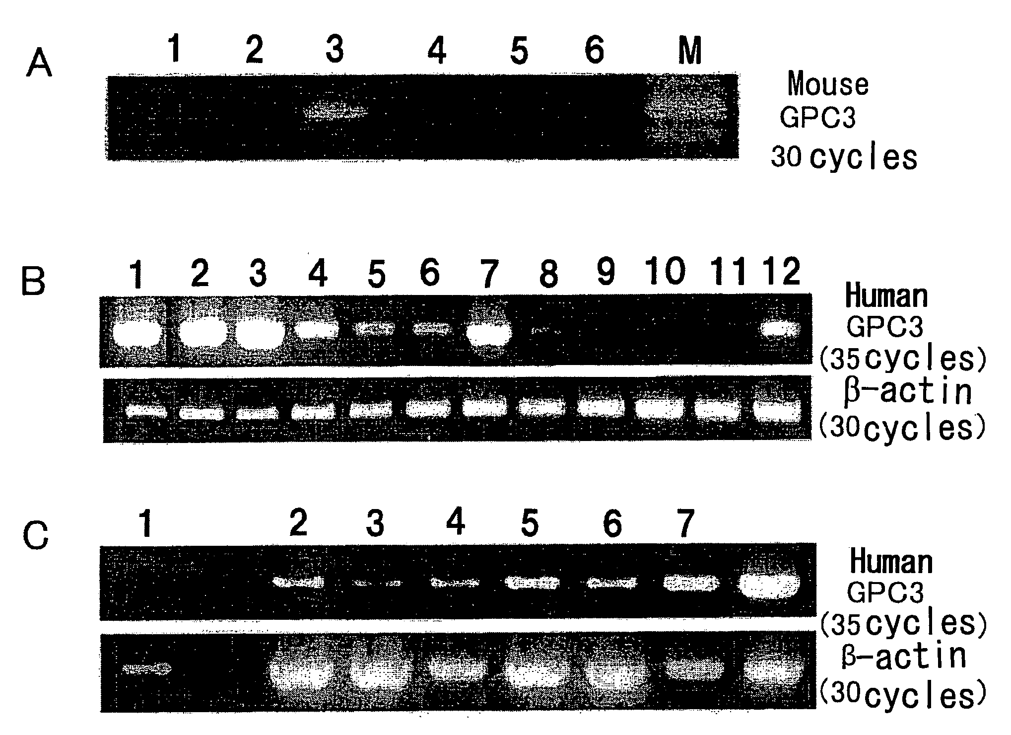
GPC3 mRNA Expression in Mouse Cell Lines

[0050]GPC3 mRNA expression was examined by reverse transcriptase-PCR (RT-PCR). MH129F, MH129P, and MH134 mouse cell lines were obtained from the Cell Resource Center for Biomedical Research, Institute of Development, Aging and Cancer, Tohoku University. EL4, Colon26, and B16 were donated by Dr. M. Ogawa of Kumamoto University.

[0051]RT-PCR was performed according to a known method (e.g., Nakatsura T. et al., Biochem. Biophys. Res. Commun. 281, 936-944 (2001)). Mouse GPC3 gene-specific PCR primers capable of amplifying a 500-bp fragment were designed. RT-PCR reaction was performed using the primers, and it consisted of 5 minutes of initial denaturation at 94° C. followed by 30 amplification cycles at an annealing temperature of 58° C. The GPC3 PCR primer sequences used herein were sense:

sense:5′-ACGGGATGGTGAAAGTGAAGA-3′(SEQ ID NO: 1)andantisense:5′-GAAAGAGAAAAGAGGGAAACA-3′.(SEQ ID NO: 2)

[0052]The mouse cell lines were compared in terms ...

Example

Example 2

GPC3 mRNA Expression in Human Melanoma Cell Lines

[0053]PC3 mRNA expression was examined by reverse transcriptase-PCR (RT-PCR). G361, CRL1579, SK-MEL-28, HMV-I, and HMV-II melanoma cell lines were obtained from the Cell Resource Center for Biomedical Research, Institute of Development, Aging and Cancer, Tohoku University. 526mel and 888mel were donated by Dr. Y. Kawakami of Keio University. Moreover, Ihara, MeWo, and colo38 were donated by Dr. T. Kageshita of Kumamoto University. Furthermore, cultured human epidermal melanocytes, HEMn, were purchased from KURABO (KURABO INDUSTRIES LTD.).

[0054]RT-PCR was performed according to a known method (e.g., Nakatsura T. et al., Biochem. Biophys. Res. Commun. 281, 936-944 (2001)). Human GPC3 gene-specific PCR primers capable of amplifying a 939-bp fragment were designed. RT-PCR reaction was performed using the primers, and it consisted of 5 minutes of initial denaturation at 94° C. followed by 30 amplification cycles at an annealing te...

Example

Example 3

GPC3 mRNA Expression in Human Melanoma Tissues

[0056]Similarly, GPC3 mRNA expression in normal human skin, human melanoma, and human pigmented nevus tissues was examined. Specimens used herein were donated by Dr. T. Kageshita, for which informed consent had been obtained from donors treated at the Department of Dermatology, Kumamoto University School of Medicine. As a result, no GPC3 mRNA expression was observed in normal skin, but it was observed in most melanoma tissues. Furthermore, GPC3 mRNA expression was also observed in congenital pigmented nevus.


PUM


Abstract

An object of the present invention is to provide a novel and clinically useful diagnostic agent for malignant melanoma. The present invention provides a diagnostic agent for malignant melanoma, which comprises an antibody against GPC3, or a probe or a primer capable of detecting GPC3 expression.

Description

TECHNICAL FIELD[0001]The present invention relates to a diagnostic agent and a diagnostic kit for malignant melanoma and a diagnostic method for malignant melanoma.BACKGROUND ART[0002]Melanoma is one type of skin cancer referred to as malignant melanoma. There are various types of skin cancer. Melanoma is the type of skin cancer that has the highest grade of malignancy. Among cells composing skin, melanin-pigment-producing cells are referred to as pigment-producing cells (melanocytes). These cells become cancerous and melanoma is developed.[0003]The frequency of the occurrence of melanoma in Japan is approximately 1.5 to 2 people per population of 100,000. It is inferred that melanoma annually occurs among approximately 1,500 to 2,000 people in Japan. In the Europe and the United States, the frequency of melanoma incidence is said to be over a dozen people per population of 100,000. The frequency of melanoma incidence in Australia is 20 or more people per population of 100,000, whic...

Claims


Application Information

Patent Timeline
IPC IPC(8): C12Q1/68A61BG01N33/574
CPCG01N33/5743
Inventor NISHIMURA, YASUHARUNAKATSURA, TETSUYA
Owner LSIP
Why Eureka
Try Eureka