
Apparatus and method for detecting lung cancer using exhaled breath

Inactive Publication Date: 2008-02-28
THE BOARD OF TRUSTEES OF THE UNIV OF ILLINOIS +1
View PDF2 Cites 34 Cited by
  • Summary
  • Abstract
  • Description
  • Claims
  • Application Information

AI Technical Summary

Benefits of technology

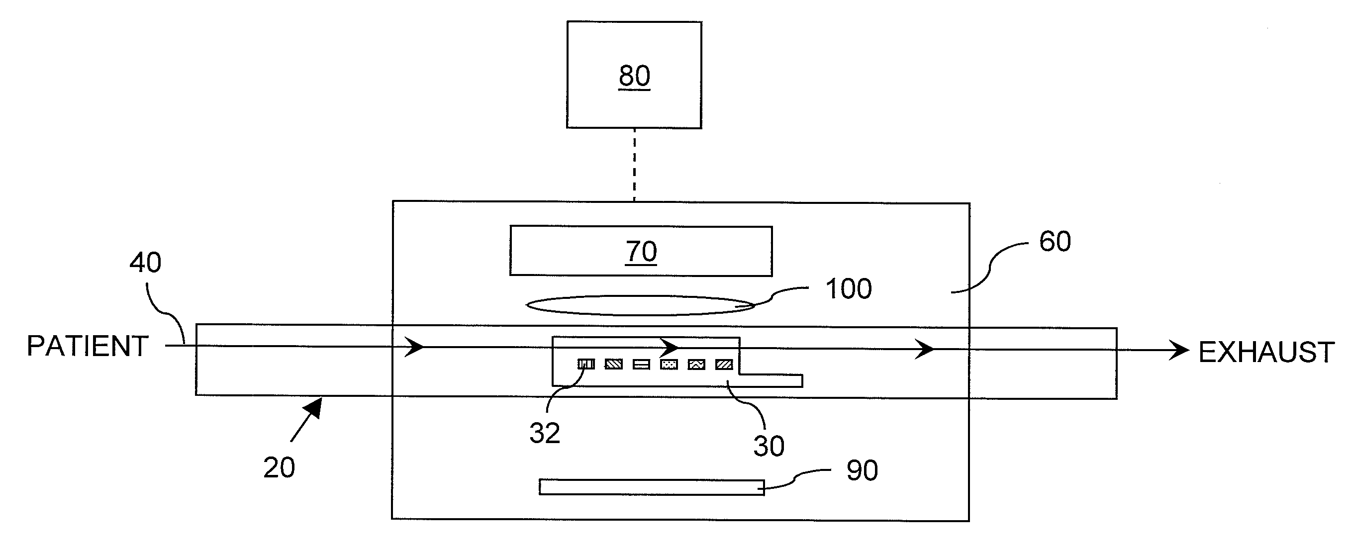
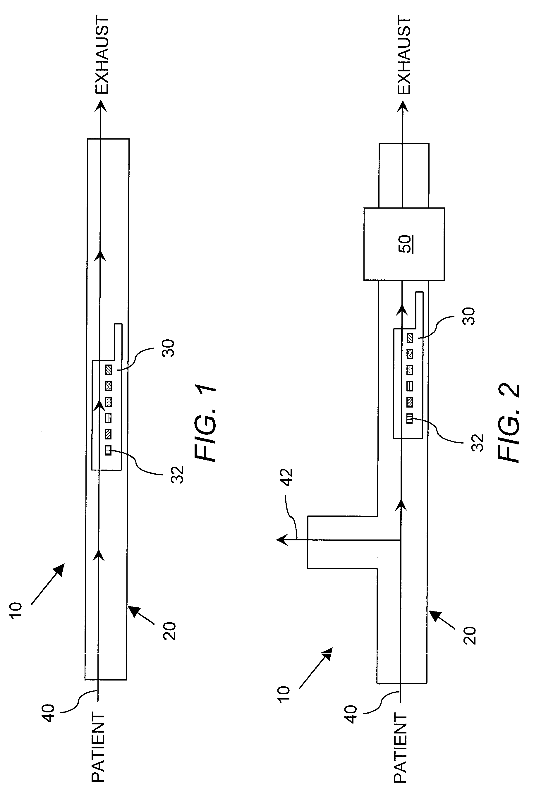
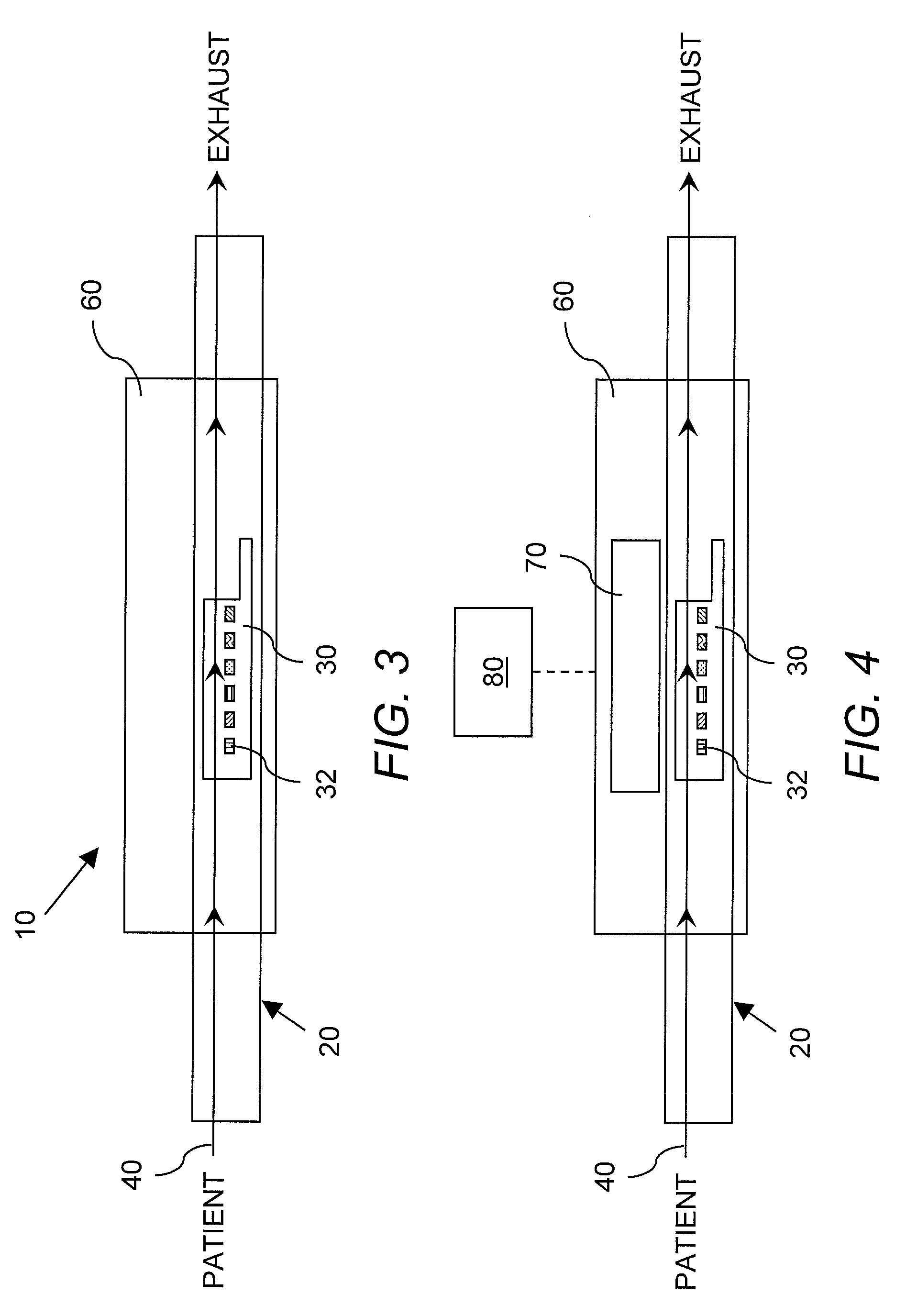
[0011] The present invention is an apparatus for detecting lung cancer. The apparatus is composed of a breath capture device including at least one colorimetric sensor array having a plurality of chemoresponsive dyes deposited thereon in a predetermined pattern combination, wherein at least one of the chemoresponsive dyes is a selected Lewis acid/base dye and wherein, in response to lung cancer analytes in exhaled breath, a distinct and direct spectral, transmission or reflectance response is produced by t

Problems solved by technology

Diagnosis of lung cancer is problematic, particularly in the early stages when it manifests no outward symptoms.
When a correct diagnosis for lung cancer is made, therefore, the cancer is often at an advanced stage, significantly reducing the likelihood of successful treatment (Jemal, et al.
Current techniques for the diagnosis of lung cancer rely on costly equipment which have the potential for complications.
Lung imaging techniques are advancing rapidly but tend to reveal multiple features, such as nodules, that can not unequivocally be diagnosed as cancer, thus requiring repeated and costly testing (Fischbach, et al.
The list of biomarkers reported for lung cancer can not be considered definitive or exhaustive, however, as the techniques used in those studies inherently favor the detection of certain analytes relative to others.
However, the sensors disclosed in these prior art references do not provide as broad a diversity of interactions with analytes as is desirable, but rather tend to exploit the weakest and least specific of intermolecular interactions, primarily van der Waals and physical adsorption interactions between sensor and analyte.
As such, both sensitivity for detection of compounds at low concentrations relative to their vapor pressures and selectivity for discrimination between compounds is compromised with these prior art sensors.
Cross-responsive sensors have seen limited application to the diagnosis of lung cancer via exhaled analytes.
The first system, however, employed only porphyrinic sensors and the latter employed only polymeric sensors, thus limiting the range of exhaled analytes that could be detected by each of these array sensors.

Method used


Image

  • Apparatus and method for detecting lung cancer using exhaled breath
  • Apparatus and method for detecting lung cancer using exhaled breath
  • Apparatus and method for detecting lung cancer using exhaled breath

Examples


example 1

Lung Cancer Detection

[0050] One hundred and forty-three individuals were examined for lung cancer both with conventional diagnostic techniques and by application of a colorimetric sensor array as disclosed herein to analyze exhaled breath. The lung cancer and lung disease status, as determined by conventional diagnostic techniques, of these individuals was listed in Table 1.

TABLE 1Disease StateNumberNon-Small Cell Carcinoma (NSCCA)49Healthy21Sarcoidosis20Pulmonary Arterial Hypertension (PAH)20Idiopathic Pulmonary Fibrosis (IPF)15Chronic Obstructive Pulmonary Disease COPD18

[0051] Subjects performed tidal breathing for 12 minutes. The breath was pulled over the colorimetric sensor array, which was positioned on a flatbed scanner, at a controlled flow rate by a downstream air pump. The entire system, to include breath capture tubing, was held at physiological temperature. Software was used to drive the scanners, collect images, analyze images, and calculate color change values for e...

example 2

Dye—Analyte Pairs

[0053] Table 3 provides a list of dyes, the analytes which the dyes can detect, and the resulting color change.

TABLE 3DyeAnalyteColor ChangeCresol Red (basic)Carbon dioxideViolet -> YellowPhenol Red (basic)Carbon dioxideRed -> YellowBromocresol GreenAmmoniaYellow -> GreenYellow -> BlueReichardt's DyeAcetic AcidBlue ->ColorlessTetraphenylporphirinatoEthanolGreen -> Brownmanganese (III) chloride[MnTPPCl]TetraphenylporphirinatoPyridineRed -> Greencobalt (III) chloride[CoTPPCl]Zinc tetraphenylorphyrinMethyl amineMaroon -> Brown[ZnTPP]TetraphenylporphyrinHydrogenBrown -> Green[H2TPP]chlorideTetraphenylporphyrinAmmoniaGreen -> Brown[H4+2TPP] (diprotonated) Bismuth (III)Hydrogen SulfideColorless ->neodecanoateBlackTetra(2,6-Hydrogen CyanideBrown -> Greendihydroxy)phenylporphyrin(with HgBr2)Copper(II)Hydrogen SulfideSky blue ->acetylacetonateBrownCopper(II)MethanethiolSky blue ->acetylacetonateBrownPalladium(II) acetateMethanethiolLight yellow ->Dark YellowPalladium(II) ...


PUM


Abstract

The present invention is an apparatus and method for detecting lung cancer. The apparatus is composed of a breath capture device including a colorimetric sensor array with a plurality of chemoresponsive dyes deposited thereon in a predetermined pattern combination, wherein the dyes produce a distinct and direct spectral, transmission or reflectance response in the presence of analytes in the exhaled breath of lung cancer patients. Air flow, temperature regulation, and visual imaging components of the instant apparatus are also provided.

Description

INTRODUCTION [0001] This application is a continuation-in-part application of U.S. patent application Ser. No. 11 / 058,497, filed Feb. 15, 2005, which is a continuation-in-part application of U.S. patent application Ser. No. 10 / 279,788, filed Oct. 24, 2002, which is a continuation-in-part application of U.S. patent application Ser. No. 09 / 705,329, filed Nov. 3, 2000, now U.S. Pat. No. 6,495,102, which is a continuation-in-part application of U.S. patent application Ser. No. 09 / 532,125, filed Mar. 21, 2000, now U.S. Pat. No. 6,368,558, all of which are incorporated herein by reference in their entireties.[0002] This invention was made in the course of research sponsored by the National Institutes of Health (Grant No. R01-HL25934). The U.S. government may have certain rights in this invention.BACKGROUND OF THE INVENTION [0003] Lung cancer causes more than 150,000 deaths in the United States each year (Patz, et al. (2004) J. Clin. Oncol. 22:2202-2206). Diagnosis of lung cancer is proble...

Claims


Application Information

Patent Timeline
IPC IPC(8): G01J3/46
CPCA61B5/083G01N21/78G01N31/22A61B5/7267G01N33/52G01N2800/12G01N33/497
Inventor SUSLICK, KENNETHMCNAMARA, WILLIAM
Owner THE BOARD OF TRUSTEES OF THE UNIV OF ILLINOIS
Learn More