Method and medical imaging system for acquisition of image data

a medical imaging and image data technology, applied in the field of medical imaging system for image data acquisition, can solve the problems of complicated situation, motion of the beating heart, and often significant imprecision

Inactive Publication Date: 2008-03-13
SIEMENS HEALTHCARE GMBH
View PDF4 Cites 7 Cited by
  • Summary
  • Abstract
  • Description
  • Claims
  • Application Information

AI Technical Summary

Benefits of technology

[0019]The inventive method also permits the reconstruction of several 3D image data records that represent the body part in several consecutive heart phases. As a result this can also be displayed dynamically. Any other images acquired with another medical imaging procedure can also be dynamically displayed in the same way, thereby creating a well synchronized 4D display of images of the heart using different imaging procedures.

Problems solved by technology

As a result, significant imprecisions often arise, because e.g. the left atrium may change.
Another problem with imaging the heart in general is the motion of the beating heart.
This situation is complicated even further because patients often do not have a regular heartbeat.

Method used


Image

  • Method and medical imaging system for acquisition of image data
  • Method and medical imaging system for acquisition of image data

Examples


Embodiment Construction

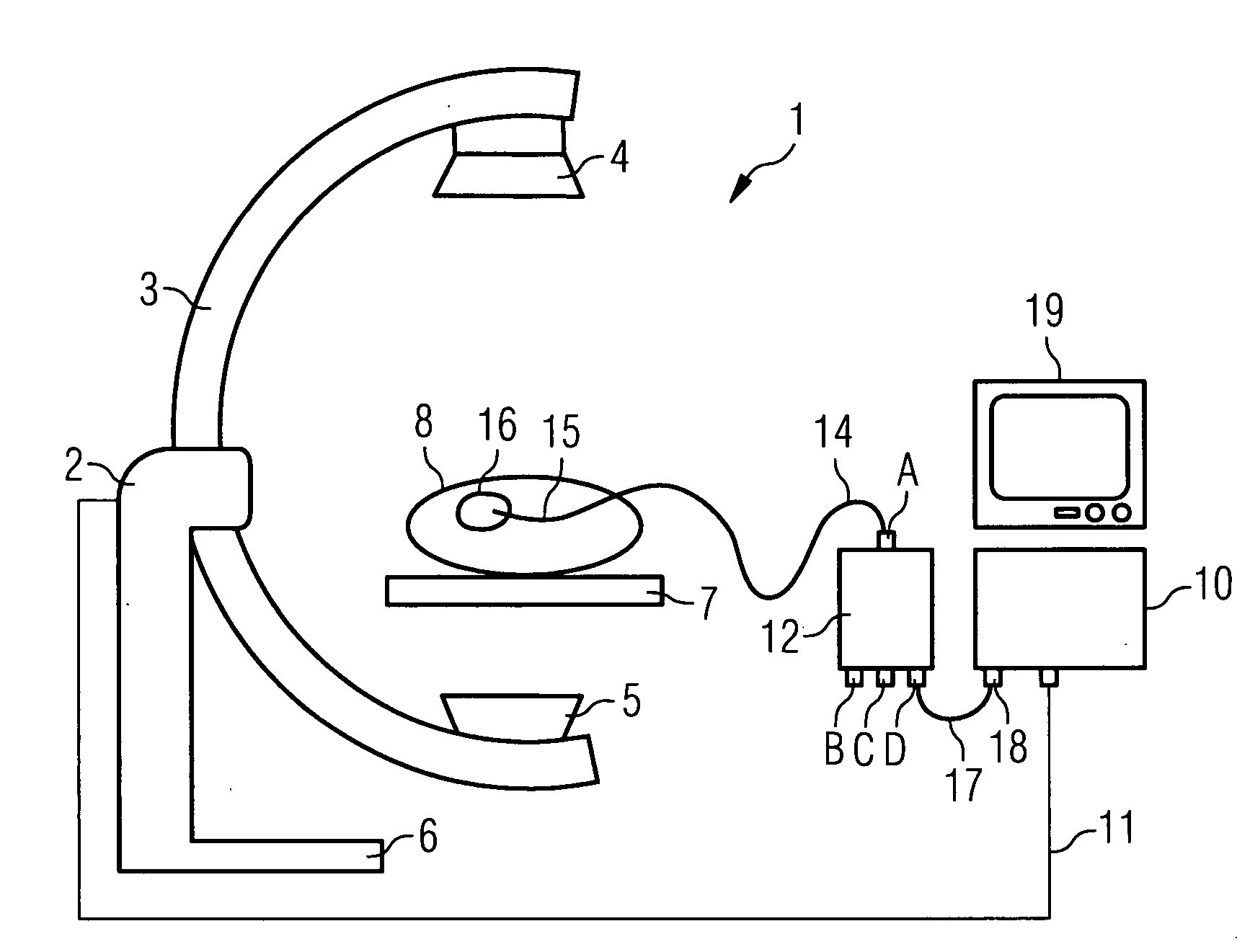
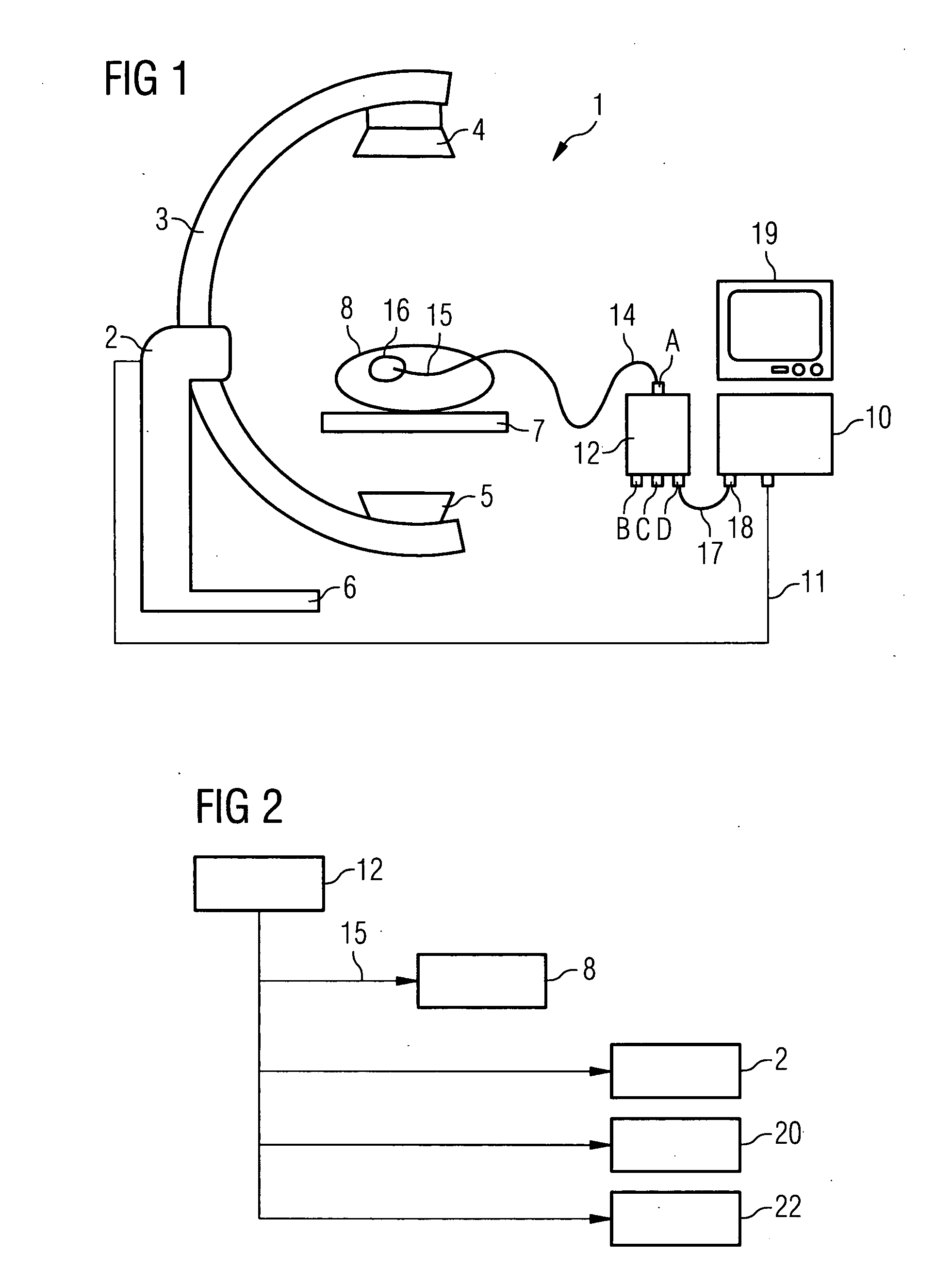
[0027]With reference to FIG. 1, an imaging system 1 is shown there which includes a C-arm x-ray device 2. This has a C-arm 3, attached to the ends of which are an x-ray source 4 and an x-ray detector 5 respectively. The C-arm 3 is held by a stand 6 and can be swiveled, especially rotated, around a patient table 7. A patient 8 is shown on the table who is to undergo a cardiological intervention. Intracardial electrodes 15 are hence inserted into the heart 16, and are connected via a cable 14 to an external heart pacemaker 12. This has outputs A, B, C and D, at which in each case the pacing signal is emitted. Output D is connected via a cable 17 to the input 18 of a control apparatus 10 of the C-arm x-ray device 2. The control unit 10 contains in particular a processor and data memory and is used to control the movements, image recording and reconstructions of the C-arm device 2. To this end it is connected to the C-arm device 2 via signal lines 11. The images are displayed for the op...


PUM


Abstract

The invention relates to a method and a medical imaging system for acquisition of image data of the heart using a medical imaging procedure during an intervention on the heart, while the heart is stimulated by a pacing signal from an external heart pacemaker, the acquisition and/or reconstruction of the image data being controlled, in particular triggered, by the pacing signal.

Description

CROSS REFERENCE TO RELATED APPLICATIONS[0001]This application claims priority of German application No. 10 2006 042 997.4 filed Sep. 13, 2006, which is incorporated by reference herein in its entirety.FIELD OF THE INVENTION[0002]The invention relates to a method for acquisition of image data of a body part of a patient using a medical imaging procedure, and a corresponding medical imaging system.BACKGROUND OF THE INVENTION[0003]In cardiology image-controlled diagnostic or therapeutic inventions are often performed on the heart, for example catheter ablation, balloon dilation and the insertion of a stent. In ablation treatments the heart wall is selectively sclerosed at selected points in order to remove electrical short-circuit paths in the heart and tachycardias associated with them. In such interventions intracardial electrodes are generally first inserted into the heart, with which the heart is stimulated by an external pacemaker during the intervention. This is called “pacing” a...

Claims


Application Information

Patent Timeline
Patent Type & Authority Applications(United States)
IPC IPC(8): A61B5/0402
CPCA61B5/0402A61B6/541A61B6/4441A61B5/318
Inventor BOESE, JANMEYER, ANDREASRAHN, NORBERT
Owner SIEMENS HEALTHCARE GMBH
Why Eureka
Try Eureka