Single-photon emission computed tomography (SPECT) using helical scanning with multiplexing multi-pinhole apertures

a multi-pinhole technology, applied in the field of single-photon emission computed tomography (spect) using helical scanning with multiplexing multi-pinhole apertures, can solve the problems of image artifacts, decrease in reconstruction quality, image artifacts, etc., to improve sensitivity and resolution in the spect imaging system, increase overlapping projections, and increase angular sampling and pinholes

Inactive Publication Date: 2008-04-17
BIOSCAN
View PDF3 Cites 8 Cited by
  • Summary
  • Abstract
  • Description
  • Claims
  • Application Information

AI Technical Summary

Benefits of technology

[0007] To improve sensitivity and resolution in SPECT imaging system a helical scan is implemented allowing an increase in overlapping projections along the axial direction of a detector. The SPECT imaging system of the present invention acquires data for an object by performing a helical scan of the object. The helical scanning of an object by a SPECT imaging system allows for artifact-free image reconstruction of said object. In addition to increased angular sampling and pinholes. These overlapped regions on the detector can potentially create null space or singularities in the imaging system and in turn result in a reconstruction of the image with artifacts. FIG. 1B shows the projections created by various multiplexing multi-pinhole aperture configurations with different percentages of overlap on the detector 102. The percentages of overlap are calculated as the percentage of overlap relative to the total area of the projections taken individually. FIG. 1C is a schematic diagram representing the effect of overlap on image reconstruction quality. The mean-squared error (MSE) between a true object and a reconstructed object is plotted as a function of the different overlap sequences presented in FIG. 1B. FIG. 1C demonstrates that as the percentage of overlap increases on a detector, the amount of artifacts introduced to a reconstructed image (mean-squared error) also increases.

Problems solved by technology

However, the overlapping projections created by the pinholes of these high-resolution, high-sensitivity SPECT imaging devices introduce sampling singularities which in turn can result in image artifacts.
The existence of these object dependent null components in turn lead to a decrease in reconstruction quality and in some cases image artifacts (FIGS. 1 & 2).
A reconstruction artifact is an impurity in the reconstructed image caused by one of a variety of effects, e.g. poor system modeling, detector failure, a large amounts of activity outside the field of view, null component in the imaging system, etc.

Method used


Image

  • Single-photon emission computed tomography (SPECT) using helical scanning with multiplexing multi-pinhole apertures
  • Single-photon emission computed tomography (SPECT) using helical scanning with multiplexing multi-pinhole apertures
  • Single-photon emission computed tomography (SPECT) using helical scanning with multiplexing multi-pinhole apertures

Examples


Embodiment Construction

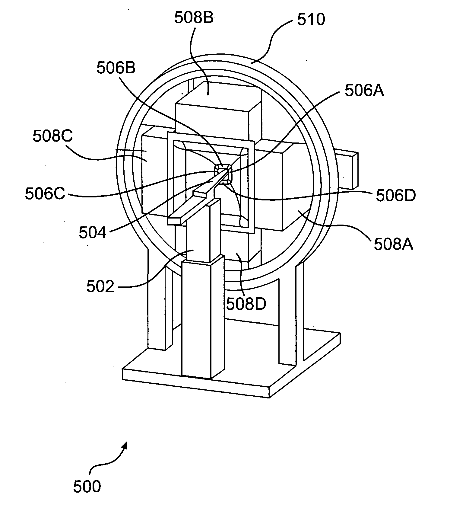
[0022] An exemplary SPECT imaging device 300, in which the present invention may be implemented, is shown in FIG. 3A. System 300 includes at least one imager 302, object support structure 304 (translation stage), and system 306. Turning here briefly to FIG. 3B. The imager 302 includes a multi-pinhole collimator 308 and a detector 310. The imager 302 is operable to rotate in the transaxial plane around an object of interest (not shown) being supported by the object support structure 304. The imager 302 can be rotated by a motor, such as a gantry, under the control of system 306. The imager 302 may be rotated a number of degrees after a projection is taken. The imager 302 can be rotated an unlimited amount either using slip-ring electronics or by using a technique by which the imager is rotated in an alternating clock-wise and counter-clockwise motion while translating the object support structure forward or backward respectively to generate a helical orbit.

[0023] In an embodiment of...


PUM


Abstract

The reconstruction of artifact free images is made possible by the implementation of a SPECT imaging device that employs helical scanning. The SPECT imaging device includes a detector configured to detect photons, such as photons, that are projected onto it. A collimator is axially aligned with the detector and includes a plurality of pinholes configured to create overlapping projections of the photons. An object support structure is configured to move in a direction that is axially aligned with the detector and collimator. The detector and collimator are configured to rotate around the object support structure in a transaxial plane to the object support structure while the object support structure moves in an axial direct to the collimator and detector.

Description

FIELD OF THE INVENTION [0001] The present invention relates to SPECT imaging using multi-pinhole apertures with overlapping projections from each pinhole in the transaxial and axial directions. BACKGROUND OF THE INVENTION [0002] SPECT technology is used in the medical field for performing such tasks as animal research, preclinical research, and patient diagnosis. Typically, radioisotopes are administered to an object of interest, such as an animal or human. The administered radioisotopes emit energy in the form of radiation that can be detected. The spatial distributions of the radioisotopes in the object of interest can be determined from the detected radioisotopes. Based on the distribution of the radioisotopes in the object, various diagnoses can be made about the object. [0003] Various types of SPECT imaging devices have been developed for the purpose of detecting radioisotopes administered to an object. The most recent SPECT imaging devices implement a multiplexing multi-pinhol...

Claims


Application Information

Patent Timeline
Patent Type & Authority Applications(United States)
IPC IPC(8): G01T1/00
CPCA61B6/027G01T1/1648
Inventor HOPPIN, JOHNVANCAUTER, STAFLACKAS, CHRISTIANNAGY, LASZLOENGELAND, UWE
Owner BIOSCAN
Why Eureka
Try Eureka