Optical surgical device and methods of use

a surgical device and optical technology, applied in the field of optical surgical devices and methods for imaging body tissue, can solve the problems of limited application, high cost of colposcope and clinical training required to perform colposcope, and inability to use, so as to reduce the risk of perforation

Inactive Publication Date: 2008-05-08
FEMSUITE
View PDF5 Cites 197 Cited by
  • Summary
  • Abstract
  • Description
  • Claims
  • Application Information

AI Technical Summary

Benefits of technology

[0016] Some embodiments of the present invention provide an optical device for inspecting body tissue. In some embodiments, the apparatus

Problems solved by technology

Colposcopy is an expensive procedure that requires a dedicated instrument, the colposcope, an a specially trained clinician.
While colposcopy is considered a preferred procedure for diagnosing cervical abnormalities, it also has come drawbacks.
The cost of the colposcope and of the clinical training required to perform it limit application.
Additionally, the colposcope is a bulky instrument, usable only in dedicated clinical settings, and provides no view of the uterus.
Another drawback of known hysteroscopes is limited maneuverability, due to the rigidity of the shaft that makes it difficult to maneuver the instrument within the patient's genital system.
Still another drawback relates to the use of fiber optics, which are manufactured from glass that breaks under bending stress, requiring frequent maintenance of the hysteroscope with consequent downtime and costs.
Additionally, in known hysterocopes the camera, saline channel, a

Method used


Image

  • Optical surgical device and methods of use
  • Optical surgical device and methods of use
  • Optical surgical device and methods of use

Examples


Embodiment Construction

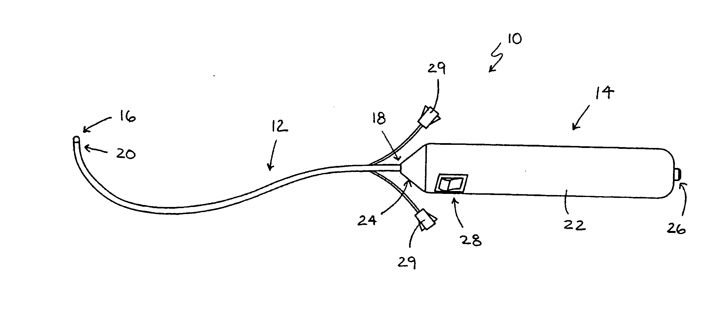
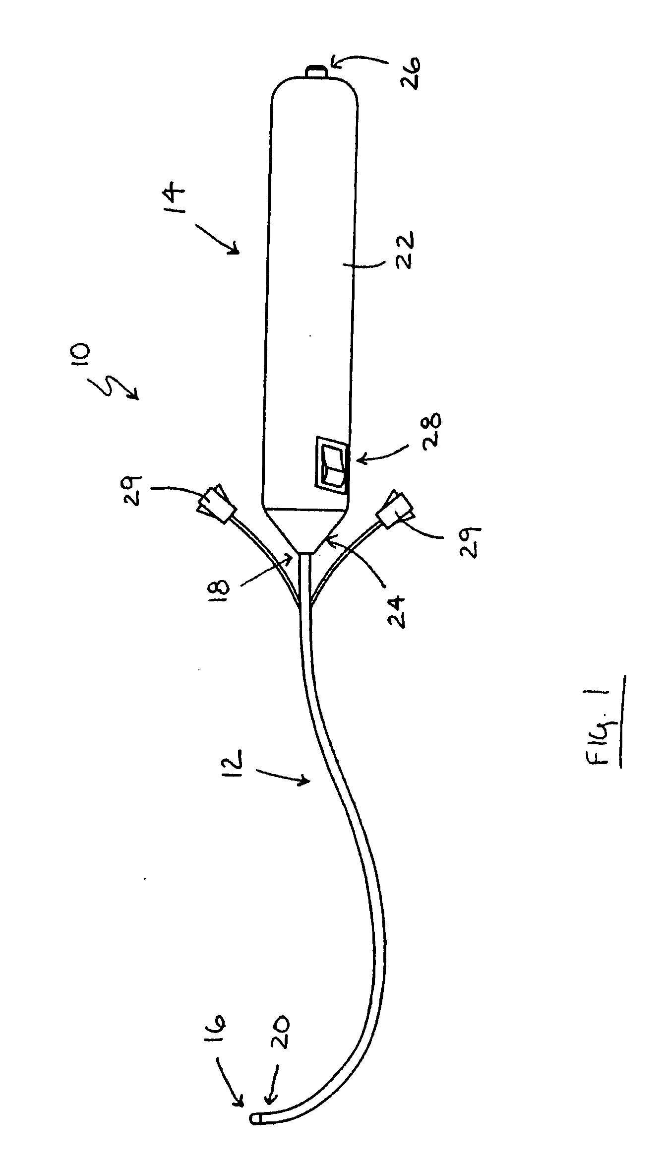
[0121] The present invention is directed to an optical device, or endoscope, for use during medical procedures to view body tissue. The device may be constructed from detachable portions that may be reused or discarded as desired so that cleaning and sterilization may be simplified.

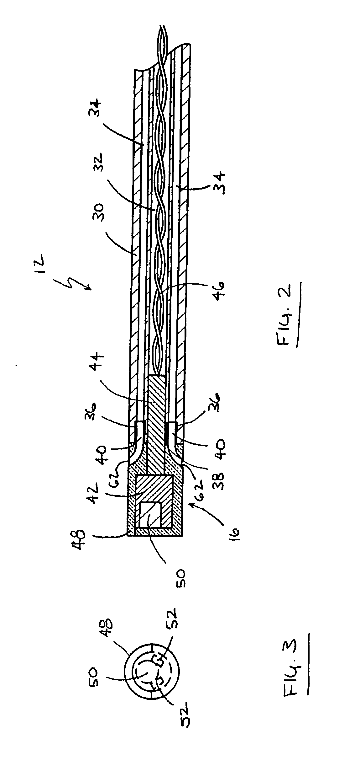
[0122] Referring to FIG. 1, an embodiment of the optical device will be described. Optical device 10 generally includes a shaft 12, handle 14 and camera assembly 16. Shaft 12 extends between handle 14 and camera assembly 16 and may be generally flexible. Shaft 12 may be configured so that after it is bent by a user it retains the bent configuration but may be easily reconfigured if desired. Such behavior is referred to herein as “shapeability,” and may be achieved by embedding stiffening members, such as stiffening wires, or blades, in the shaft as will be described in greater detail below. Fluid connectors 29 may also be provided so that fluid may be injected or aspirated through fluid conduits in shaft...


PUM


Abstract

An optical device includes a shaft, a handle and a camera assembly. The handle is coupled to the shaft at a first end, and the camera assembly is coupled to the shaft at a second end. Camera circuitry and software may be provided in the shaft and the handle, so that, in one embodiment, the device may be constructed with reusable portions of the camera circuitry and software. In another embodiment, the device may be provided as a single piece, that may be discarded or sterilized after use.

Description

CROSS-REFERENCE TO RELATED APPLICATIONS [0001] This application claims priority to U.S. provisional patent application Nos. 60 / 853,161, filed Oct. 20, 2006, 60 / 878,892, filed Jan. 4, 2007, 60 / 903,583, filed Feb. 26, 2007, 60 / 921,925, filed Apr. 4, 2007, 60 / 925,486, filed Apr. 20, 2007, and 60 / 933,233, filed Jun. 4, 2007, all of which are hereby incorporated by reference herein in their entireties.FIELD OF THE INVENTION [0002] The present invention relates to apparatus and methods for imaging body tissue during medical procedures. More particularly, the present invention relates to apparatus and methods that provide endoscopic viewing of the female genital tract during gynecological procedures. BACKGROUND OF THE INVENTION [0003] A number of gynecological procedures require visual inspection of the genital tract of a female patient, which is often performed with the use of an endoscope, or hysteroscope. Conventional endoscopes are often constructed from a rigid body and oftentimes tho...

Claims


Application Information

Patent Timeline
IPC IPC(8): A61B1/04
CPCA61B1/00105A61B1/303A61B1/00052A61B1/00103A61B1/0676A61B1/015A61B1/018A61B1/05A61B1/00124
Inventor SANDERS, GERALD J.SIKRI, VIVEK
Owner FEMSUITE
Why Eureka
Try Eureka