

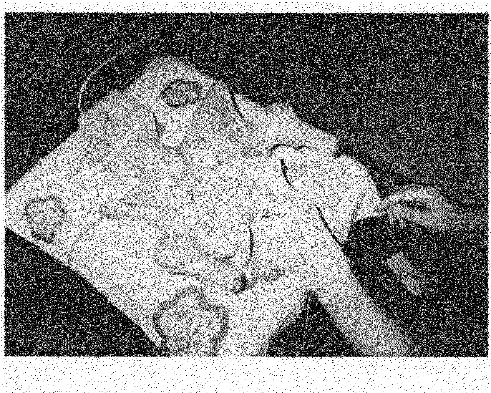
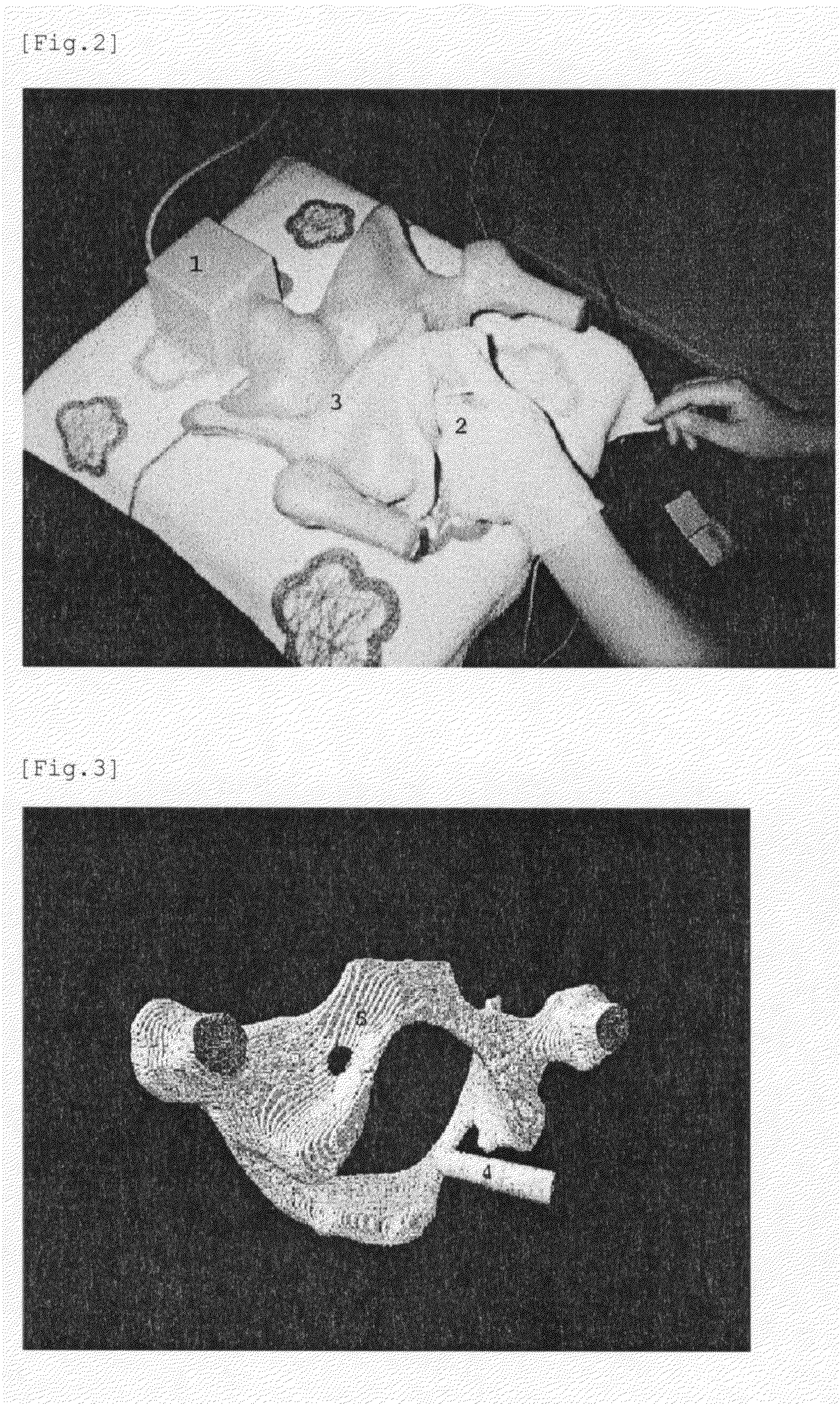
Medical Training Model Device
a technology of medical training and model device, applied in the field of teaching material model system, can solve the problems of no appropriate model, difficult to grasp, and practice internal examination
- Summary
- Abstract
- Description
- Claims
- Application Information
AI Technical Summary
Benefits of technology
Problems solved by technology
Method used
Image

Examples
example
[0040]When the system of the present invention is practiced, the flow of data processing is as follows.
1) Preparation of Three-Dimensional Model Data (the Pelvis Data etc.)
[0041]In the present system, three-dimensional data of the teaching material model being the actual teaching material model for clinical examination inside of an organism are previously prepared, which are displayed on the display screen creating means as three-dimensional CG. For the data of actual teaching material model, in the case of an organism, from a three-dimensional image imported through CT scan or MRI, a three-dimensional model, that is, three-dimensional CG is established by using equivalent-face processing or segmentation technique. The teaching material model is previously modeled by using a geometric modeling software.
2) Preparation of Three-Dimensional Data for the Figure of the Intra-Organ Inserting Means (Internal Examination Finger, Endoscope etc.)
[0042]The data for the inserting means (hereina...
PUM
 Abstract
Description
Claims
Application Information