Pre-And Intra-Operative Imaging of Testicular Torsion
a testicular and intraoperative technology, applied in the field of preand intraoperative imaging of testicular torsion, can solve the problems of high cost, time-consuming, and difficult to achieve, and achieve the effect of facilitating such determinations
- Summary
- Abstract
- Description
- Claims
- Application Information
AI Technical Summary
Benefits of technology
Problems solved by technology
Method used
Image
Examples
example 1
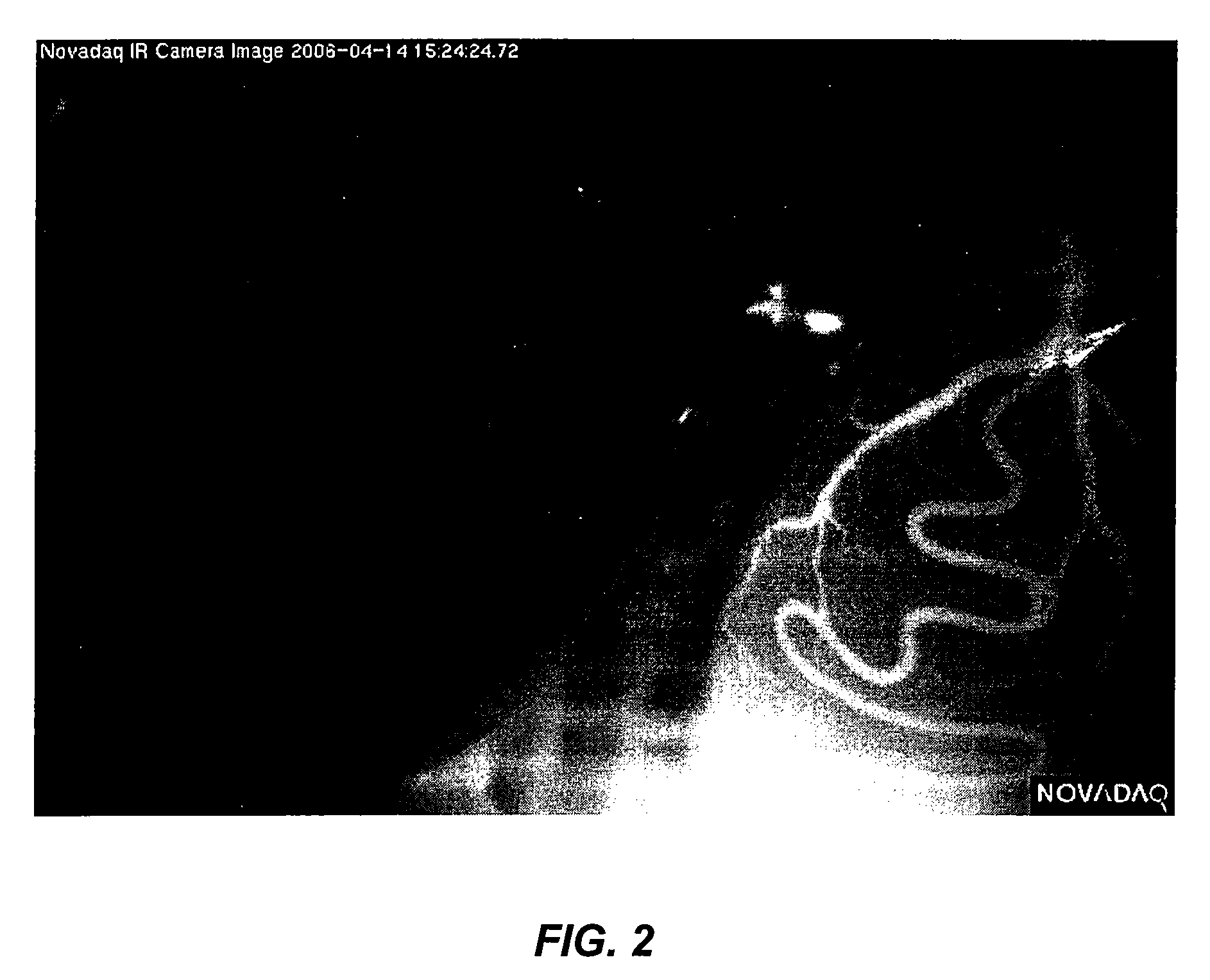
[0047]Intraoperative video angiography is performed with a laser-fluorescence imaging device (Novadaq Technologies, Inc., Toronto, Canada) consisting of a near infrared (NIR) laser light source and a NIR-sensitive digital camcorder. For measurements, the unit is positioned 30 to 40 cm from the area of interest. ICG, dissolved in water, is then injected as a bolus. When ICG is used as the imaging dye, NIR light emitted by the laser light source induces ICG fluorescence. The fluorescence is typically imaged by a video camera, with optical filtering to block ambient and laser light so that only ICG fluorescence is captured. Images can be viewed by the surgical team on screen in real time (typically 25 images / sec). Optionally, the images can be stored on the video camera or transferred to a computer or to storage media for later review or training of others.
example 2
[0048]Two rats were subjected to unilateral interruption of blood flow to the right testis. In rat A, a bulldog clamp was used, in rat B, a mosquito clamp (a smaller clamp designed to cut off blood flow in small blood vessels) was used. ICG was administered by IV to each rat to determine the ability to visualize the degree to which the testicle underwent hypoperfusion or lack of perfusion.
[0049]Prior to administration of ICG, rat A was first subjected to near IR and imaged. No testicular fluorescence was evident prior to ICG administration. 0.5 ml of ICG at a concentration of 2.5 mg / ml was then administered by IV. The right spermatic cord was clamped with a bulldog clamp to create vascular obstruction and the testicles were imaged. There was low fluorescence in the right hemiscrotum and intense fluorescence in the left hemiscrotum. Minimal fluorescence in the right hemiscrotum appeared due to the presence of ICG in the subcutaneous and cutaneous vessels of the scrotum; the different...
PUM
 Abstract
Description
Claims
Application Information
 - R&D
- Intellectual Property
- Life Sciences
- Materials
- Tech Scout
- Unparalleled Data Quality
- Higher Quality Content
- 60% Fewer Hallucinations
Browse by: Latest US Patents, China's latest patents, Technical Efficacy Thesaurus, Application Domain, Technology Topic, Popular Technical Reports.
© 2025 PatSnap. All rights reserved.Legal|Privacy policy|Modern Slavery Act Transparency Statement|Sitemap|About US| Contact US: help@patsnap.com


