Device For White Balancing And Appying An Anti-Fog Agent To Medical Videoscopes Prior To Medical Procedures

a technology of white balance and camera system, which is applied in the field of white balance devices for medical videoscopes, can solve the problems of inability of digital cameras to adapt to these changes, inability of most expensive video cameras to automatically do what the eye does, and doctors do not understand the importance, so as to improve visualization and achieve accurate white balance. , the effect of easy to us

Active Publication Date: 2008-07-03
TYCO HEALTHCARE GRP LP
View PDF7 Cites 94 Cited by
  • Summary
  • Abstract
  • Description
  • Claims
  • Application Information

AI Technical Summary

Benefits of technology

[0019]Additionally, when the scope is inside the device for white balancing, the lens is making simultaneous contact with a solution or agent formulated to inhibit fog on the medical videoscope or aid in cleaning blood and debris from the lens. The liquid additionally provides an improvement in visualization. Fog is a major problem during endoscopic surgery. When cold scopes are introduced into the warm moist body, condensation occurs. This condensation fogs the viewing area forcing the procedure to be delayed until it clears up.
[0020]What is needed is a device that not only makes white balancing accurate and easy but also a device which includes a defogging mechanism. The defogging mechanism may be with the use of a cold anti-fog liquid or and electrical or exothermic mechanism that heats the anti-fog liquid. The device is opened prior to any medical procedure and placed over the distal lens of the medical videoscope. White balancing is performed by pressing the white balance button in the camera equipment. The device is then removed from the distal scope immediately prior to its insertion into the body and can be reinserted into the device whenever the scope is removed from the body. The device is sterile, for single patient use, and can be discarded after each procedure or kept on the distal scope until the scope is resterilized.

Problems solved by technology

Unlike a human eye, a digital camera is unable to adapt to these changes.
Unfortunately, even the most expensive video cameras cannot automatically do what the eye does, so we have to show our cameras what we want them to read as “white” in any given scene.
Often, doctors do not understand the importance of white balancing and so they will use a reference such as surgical gauze—which is actually full of holes and not truly white—to set the calibration.
Moreover, white balancing is carried out in the open room where there is different light sources illuminating the “white” gauze sponge; this is a problem because the video inside the body will be generated using only the camera's fiber optic light source.
Both of these mistakes generate an incorrect white balance point.
For photography, this is annoying.
For medicine this can be dangerous.
Moreover, doctors also often hold the scope too close to the white target distorting the camera's light, or too far exposing the white to room fluorescent lights and spot lights.
Another problem is that, currently, white balancing the medical videoscope during medical procedures is a hassle.
The doctor is often sterile and cannot press the white balance button on the camera equipment.
This becomes complicated and time consuming because the nurse often is busy when the surgeon is ready to white balance or the surgeon is busy when the nurse is ready to white balance.
This wastes time, and time is very expensive in the operating room.
The fogging of the scope is very costly.
When scopes fog up during surgery, the surgeon cannot see and must pause the surgery until the picture can be cleared up.
With the incredible costs relating to anesthesia and surgical staff, the wasted time in the aggregate equals hundreds and thousands of dollars.
Since many medical procedures are sterile, current methods to solve both these visualization problems are limited to messy anti-fog solutions and inaccurate white balancing techniques.
Another major problem is that the white reference must often be sterile, so doctors typically use the “white” surgical gauze sponge to white balance.
These sponges are in reality full of holes and actually not the ideal white.
Ultimately, this may distort the accuracy of the white balance and diminish the quality and the true reproduction of the color in the video generated from inside the body during the medical procedure.
Having below optimum video color quality could be dangerous as inflammation and cancer metastasis often presents as discreet color changes.

Method used


Image

  • Device For White Balancing And Appying An Anti-Fog Agent To Medical Videoscopes Prior To Medical Procedures
  • Device For White Balancing And Appying An Anti-Fog Agent To Medical Videoscopes Prior To Medical Procedures
  • Device For White Balancing And Appying An Anti-Fog Agent To Medical Videoscopes Prior To Medical Procedures

Examples


Embodiment Construction

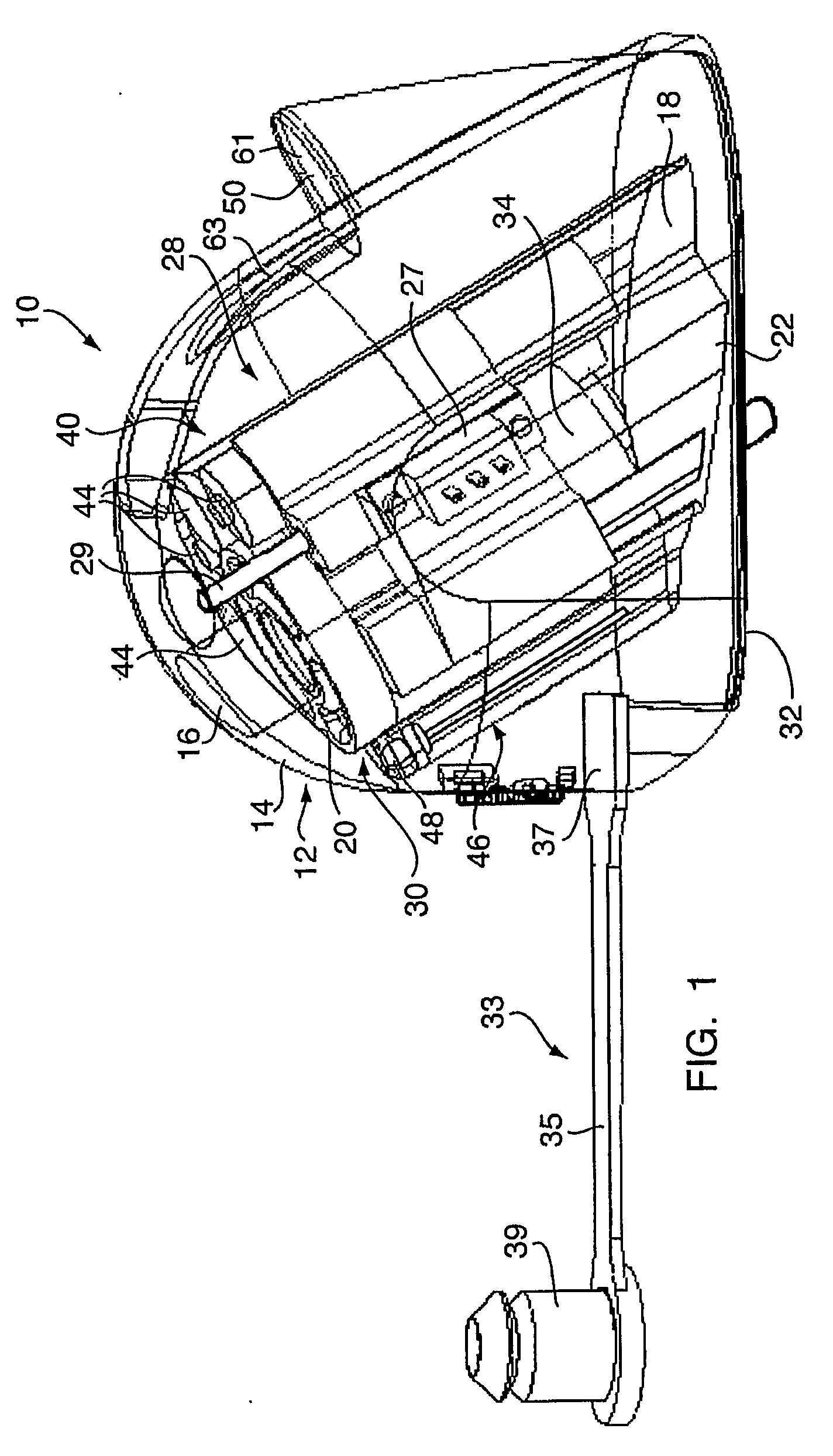
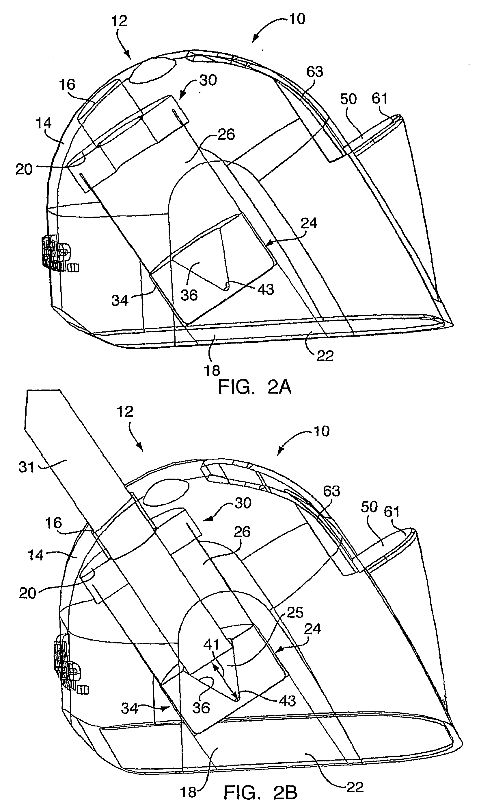
[0053]With reference to FIGS. 1 through 3F, a white balance device embodying the present invention is indicated generally by the reference number 10. The device 10 comprises a housing or outer shell 12. The housing 12 has an outer surface 14 defining an opening 16 for inserting therein a medical videoscope such as a laparoscope or endoscope. An interior of the housing 12 defines a canal 18 having a first end 20 communicating with the opening 16 and a second end 22 terminating within the housing 12 for receiving a distal lens of a medical videoscope. A white balancing reference material 24 (see FIGS. 5A through 5C) is disposed within the housing 12 adjacent to the second end 22 of the canal 18.

[0054]The device 10 preferably accommodates a defogging material 26 adjacent to the second end 22 of the canal 18 for treating and preventing the distal lens of a medical videoscope from fogging during a medical procedure. The device 10 preferably further comprises a heating mechanism 28 in the...


PUM


Abstract

A device is configured for white balancing a medical videoscopic camera system prior to videoscopic medical procedures, as well as optionally simultaneously or non-simultaneously applying a fog-prohibiting agent to the distal lens of a medical videoscope such as an endoscope or laparoscope. The device combines a white balancing mechanism, protective mechanism, and defogging mechanism in one simple easy to use device.

Description

CROSS-REFERENCE TO RELATED APPLICATION[0001]This application claims the benefit of U.S. Provisional Application No. 60 / 763,472, filed on Jan. 30, 2006, the disclosure of which is hereby incorporated by reference in its entirety.FIELD OF INVENTION[0002]This invention generally relates to a device for white balancing a camera, and more specifically relates to a device for white balancing a medical videoscopic camera system prior to videoscopic medical procedures, as well as optionally simultaneously or non-simultaneously applying a fog-prohibiting agent to the distal lens of a medical videoscope such as, for example, an endoscope, laparoscope, bronchoscope, cystoscope or otoscope. The device combines a white balancing mechanism, protective mechanism, and defogging mechanism in one simple easy to use device.BACKGROUND[0003]The color of light reflected off of a subject changes with the color of the light source. Unlike a human eye, a digital camera is unable to adapt to these changes. T...

Claims


Application Information

Patent Timeline
Patent Type & Authority Applications(United States)
IPC IPC(8): A61B1/04
CPCA61B1/00057A61B1/045A61B1/127A61B1/125A61B1/2676A61B1/313H04N9/735A61B1/227H04N23/88A61B1/00
Inventor GOMEZ, RICARDO ALEXANDER
Owner TYCO HEALTHCARE GRP LP
Why Eureka
Try Eureka