Magnetic resonance imaging apparatus for scanning the spine

a magnetic resonance imaging and spine technology, applied in the field of magnetic resonance imaging apparatus, can solve the problems of reducing patient comfort, narrow patient space, and high cost, and achieve the effects of reducing costs, increasing patient space in the emitting body coil, and simplifying the design of the patient tabl

Active Publication Date: 2008-08-28
SIEMENS HEALTHCARE GMBH
View PDF3 Cites 9 Cited by
  • Summary
  • Abstract
  • Description
  • Claims
  • Application Information

AI Technical Summary

Benefits of technology

[0013]An object of the present invention is to provide a magnetic resonance imaging apparatus for scanning a spine, in which by changing the position for fitting the spine coil the patient's space is increased so as to improve the patient's comfort, and by reducing the number of radio frequency components in the spine coil the equipment costs are reduced.

Problems solved by technology

1) the relative movement between the patient and the spine coil is impossible, so the spine coil needs to have a sufficient length in order to achieve the scan of a whole human spine, therefore the coil needs to use a large number of radio frequency component units, which lead to high costs;
2) as shown in FIG. 2, since there is the spine coil between the patient and the patient table, it leads to the reduction of the distance HI between the inner wall of the upper part of the emitting body coil and the patient, resulting in a narrow space for the patient, and reducing the patient's comfort; and
3) since there exists certain coupling between the spine coil and the emitting body coil, it is necessary to unplug the spine coil when the apparatus is not in operation and to re-plug it into the corresponding socket for next operation. Such repeated plugging and unplugging increases the probability of damaging the coil, and therefore affects the reliability of the coil and the imaging quality of the apparatus.
Currently, a common solution is to embed the spine coil into the board of the patient table, but by doing so the structural complexity of the board of the patient table is increased, and at the same time during its use the coil is still subject to factors such as the patient's body weight, etc., so the problem regarding vulnerability to damage still exists.

Method used


Image

  • Magnetic resonance imaging apparatus for scanning the spine
  • Magnetic resonance imaging apparatus for scanning the spine
  • Magnetic resonance imaging apparatus for scanning the spine

Examples


Embodiment Construction

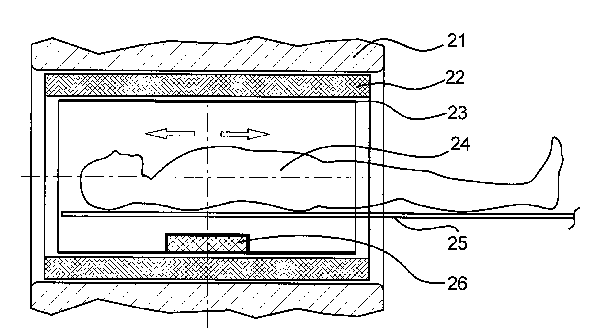
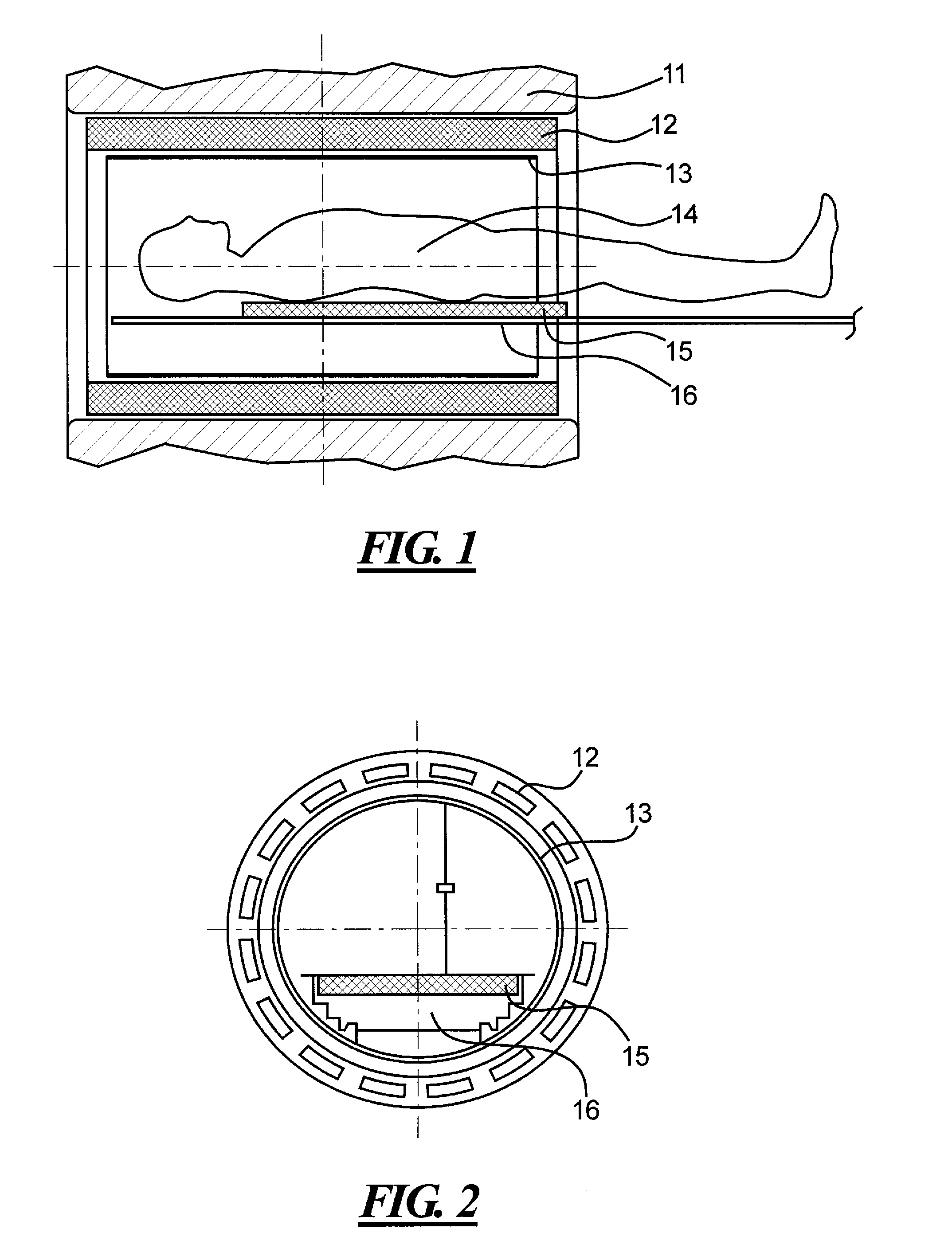
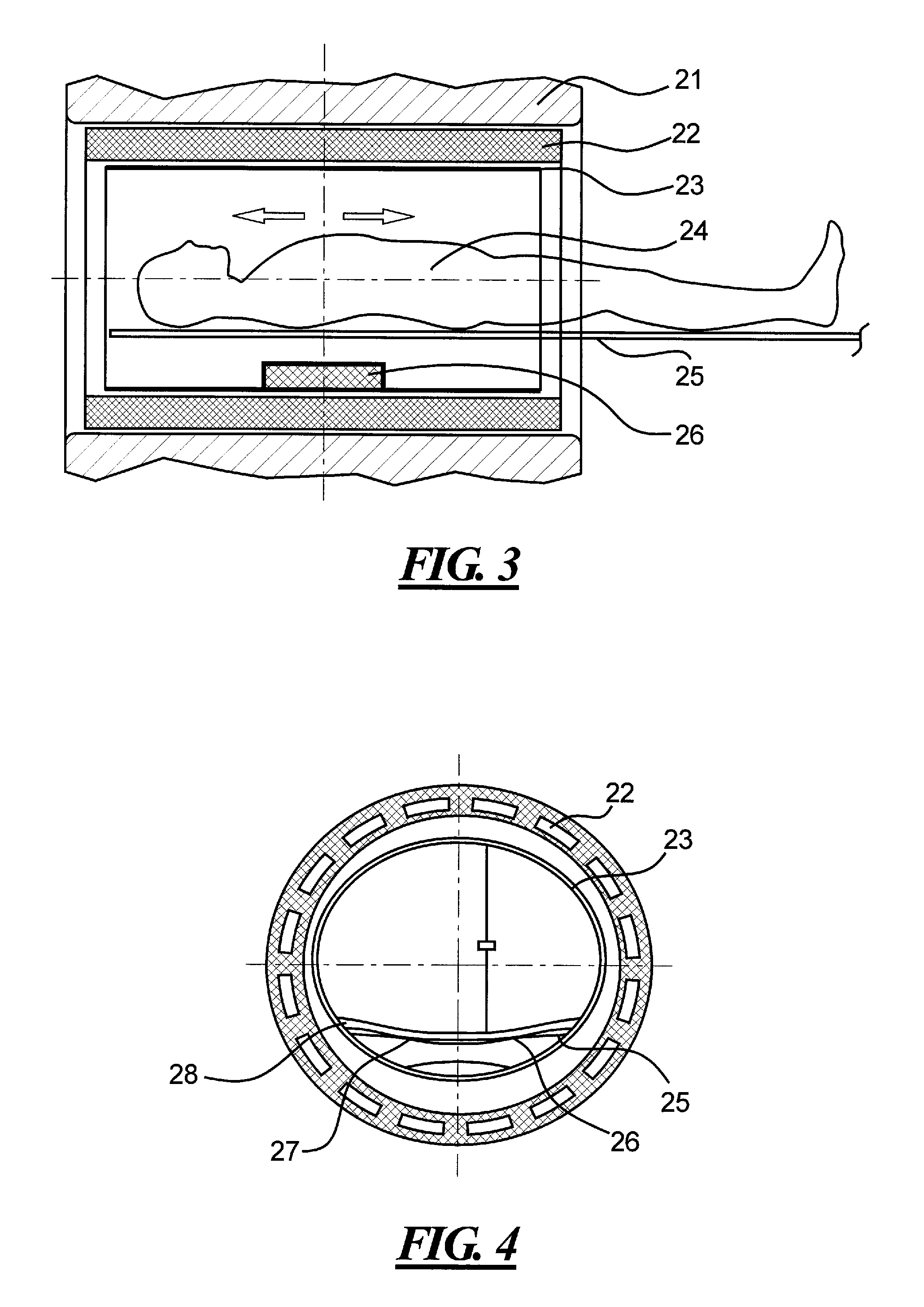
[0028]The primary basis of the present invention is to move the spine coil from the upper surface of the patient table or the interior of the board of the patient table to beneath the patient table, so as to solve the problems, such as the narrow space and high equipment costs in the prior art caused by disposing the spine coil on the upper surface of the patient table or embedding the same into the board of the patient table.

[0029]One manner for realizing the magnetic resonance imaging apparatus for scanning a spine of the present invention is to fix the spine coil directly on the lower surface of the board of the patient table. Compared with the prior art, this does not change the length of the spine coil, and it only changes the fitting position of the spine coil. However, after having fitted the spine coil to the lower surface of the board of the patient table, the distance between the inner wall of the body coil above the patient table and the patient is increased, so the patie...


PUM


Abstract

The present invention discloses a magnetic resonance imaging apparatus for scanning a spine, comprising a body coil for emitting signals, a patient table within the body coil and a spine coil for receiving signals, wherein said spine coil is fixed within said body coil and disposed under said patient table. Said patient table is movable within said body coil by slide rails fitted at the two sides thereof, so as to reduce the length of said spine coil. The cross section of said patient table is in an arched shape, and the cross section of said spine coil is in an arched shape which matches that of said patient table. By using the apparatus of the present invention, since the spine coil is disposed underneath the board of the patient table, the design and production of the board of the patient table are simplified, the number of radio frequency choke coils and radio frequency element units is reduced and so are the costs; also, the space for the patient is increased, so the patient's comfort is improved; and furthermore, the repeated plugging and unplugging of the spine coil, as in the prior art, are avoided so as to reduce the probability of damaging the coil.

Description

BACKGROUND OF THE INVENTION[0001]1. Field of the Invention[0002]The present invention relates to a magnetic resonance imaging apparatus, and particularly to a magnetic resonance imaging apparatus for scanning a spine.[0003]2. Description of the Prior Art[0004]Magnetic resonance imaging (MRI) is an imaging technology for obtaining internal structural information of an object by means of the magnetic resonance phenomena. MRI is broadly used in the field of medical imaging due to its significant advantages, such as a large number of imaging parameters, high resolution, tomography in any layer required, non-destructive imaging, etc.[0005]It is normally considered that if a surface coil is close to a patient, the image signal would be strong, the noise level low, the signal-to-noise ratio high, and the images relatively fine. When performing the magnetic resonance imaging on a human spine, a spine coil is usually used in order to obtain magnetic resonance images of relatively high qualit...

Claims


Application Information

Patent Timeline
Patent Type & Authority Applications(United States)
IPC IPC(8): G01V3/00A61B5/05
CPCA61B5/0555G01R33/307G01R33/56383G01R33/56375G01R33/341A61B5/055A61B5/704
Inventor DU, JIAN JUNKREISCHER, LUDWIGLI, JIAN ZHONGPEI, JIAN HUA
Owner SIEMENS HEALTHCARE GMBH
Why Eureka
Try Eureka