Eureka AIR delivers breakthrough ideas for toughest innovation challenges, trusted by R&D personnel around the world.

System and method for patient setup for radiotherapy treatment

Inactive Publication Date: 2008-09-11
RESONANT MEDICAL
View PDF85 Cites 24 Cited by
  • Summary
  • Abstract
  • Description
  • Claims
  • Application Information

AI Technical Summary

Benefits of technology

[0013]For deep internal organs that may require radiation treatment, such as the prostate, slight differences in the location of the region of interest within the patient from one treatment session to another can be corrected for by simply shifting the treatment couch to realign the region to its planning position. Differences and shifts in the external anatomy are of secondary importance and may have minimal effect on the required treatment plan. This is due, at least in part, to the fact that slight differences in the depth, and thus attenuation, of the radiation beam through the body are less significant when the depths are large. As a result, slight differences in the distance from the surface of the skin to the treatment region do not have a great impact on the radiation dose delivered to that region. In the treatment of deeply located organs, therefore, the value of obtaining both internal anatomical information and external information prior to every treatment session is limited. A simple repositioning of the patient may be made to compensate for anatomical changes when treating deeply located lesions.
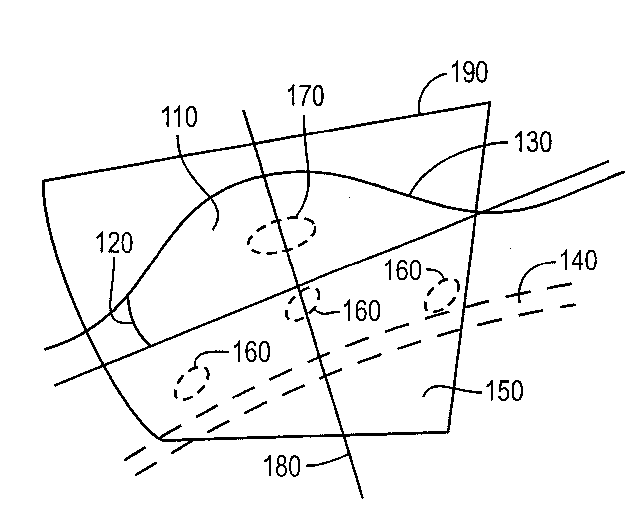
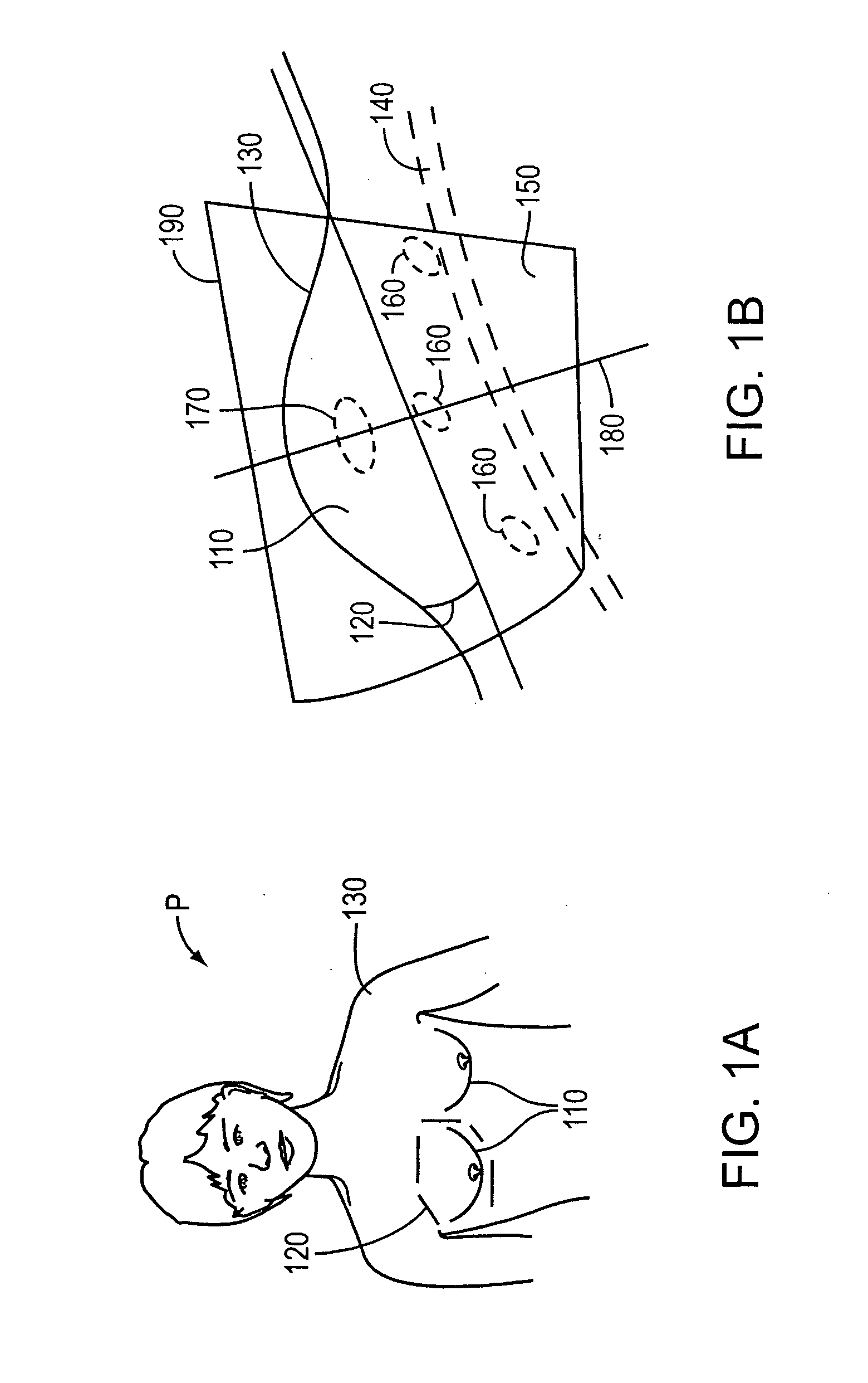
[0014]For cancerous tissue located near the surface of the skin, however, such as lesions within a patient's breast, attenuation of a radiation beam passing through this region can produce a significant change in the radiation actually received at the lesion. As a result, it is very important when treating near surface lesions to know both the location of the treatment region and the depth of this region below the surface of the skin. The present invention, by using both external information (in order to correctly locate the treatment region with respect to the patient) and internal anatomical information (to correctly measure the depth of that region below the surface), accurately corrects for morphological and conformational changes to provide the desired dose to the proper anatomical region. Thus, the approach of the present invention is especially useful when treating near-surface lesions, or lesions encompassed within a surface which can deform significantly. By contrast, prior techniques for locating breast lesions for treatment, which generally align the breast using previously created external markings alone, do not account for possible changes in the depth of the lesion below the surface of the skin.

Problems solved by technology

As a result, slight differences in the distance from the surface of the skin to the treatment region do not have a great impact on the radiation dose delivered to that region.
In the treatment of deeply located organs, therefore, the value of obtaining both internal anatomical information and external information prior to every treatment session is limited.
For cancerous tissue located near the surface of the skin, however, such as lesions within a patient's breast, attenuation of a radiation beam passing through this region can produce a significant change in the radiation actually received at the lesion.
By contrast, prior techniques for locating breast lesions for treatment, which generally align the breast using previously created external markings alone, do not account for possible changes in the depth of the lesion below the surface of the skin.
Indeed, even though conventional CT techniques reveal surface information, that information must usually be extracted using, for example, a threshold algorithm that may be inconvenient or inaccurate.
Finally, if fiducials are implanted inside a tumor, conventional projection x-rays will not provide three-dimensional surface information.
These can be detected with a set of two or more projection x-ray images which will characterize the internal anatomy and suggest how it should be placed relative to a treatment beam, but surface information cannot readily be extracted from these projection images.

Method used


Image

  • System and method for patient setup for radiotherapy treatment
  • System and method for patient setup for radiotherapy treatment
  • System and method for patient setup for radiotherapy treatment

Examples


Embodiment Construction

[0039]Throughout the following descriptions and examples, the invention is described in the context of positioning a patient in preparation for the delivery of radiation therapy to a breast. However, it is to be understood that the present invention may be applied in cases in which a patient is positioned in anticipation of receiving any position-based treatment and for any anatomical feature of the body, be it internal (e.g., a tumor within the breast surgical bed) or external (e.g., a melanoma on the skin).

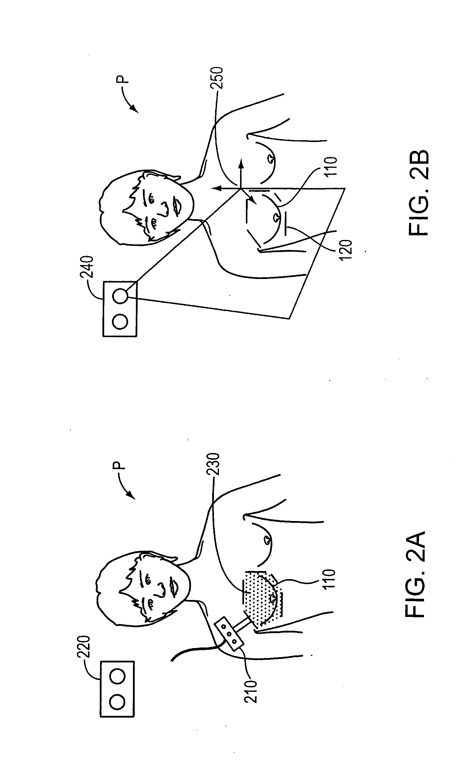
[0040]In one embodiment, the invention generally involves four phases: receiving a previously defined treatment plan, obtaining patient surface information, obtaining internal anatomical information, and correcting the treatment plan. In some embodiments, however, the treatment plan can be developed just prior to treatment, even while the patient is in the treatment room awaiting delivery of radiotherapy. Although such an approach minimizes positioning errors between the plannin...


PUM


Abstract

Positioning an anatomical feature of a patient during repeated radiotherapy treatments, and accounting for variations in that position between treatments allow a patient to be placed in a substantially repeatable orientation with respect to a treatment device.

Description

CROSS-REFERENCE TO RELATED APPLICATIONS[0001]This application claims priority to and the benefit of U.S. provisional patent application Ser. No. 60 / 714,397, filed Sep. 6, 2005, the entire disclosure of which is hereby incorporated herein by reference in its entirety.FIELD OF THE INVENTION[0002]The present invention relates generally to the field of radiotherapy, and more particularly to positioning an anatomical feature of a patient during repeated treatments, and accounting for variations in positioning between and / or during treatments.BACKGROUND OF THE INVENTION[0003]Cancerous tumors on or within an anatomical feature of a patient are often treated using radiation therapy involving one or more radiation-emitting devices. The primary goal of radiation therapy is the complete eradication of the cancerous cells, while the secondary goal is to avoid, to the maximum possible extent, damaging healthy tissue and organs in the vicinity of the tumor. Typically, a radiation therapy device i...

Claims


Application Information

Patent Timeline
IPC IPC(8): A61N5/10
CPCA61N5/1049A61N2005/1059A61N2005/1058A61B8/4254
Inventor FALCO, TONYLACHAINE, MARTINBONNEVILLE, MARTIN
Owner RESONANT MEDICAL
Why Eureka
Eureka Blog
Learn More