Method for high-resolution presentation of filigree vessel implants in angiographic images

a technology of filigree vessel and angiographic image, which is applied in the field of high-resolution presentation of filigree vessel implants in angiographic images, can solve the problems of insufficient number of verifiable quanta per detector surface from the x-ray source and the image acquired, limited power of the x-ray tube, and still poses a problem of artifact-free visualization, localization, identification and status evaluation of filigree vessel implants

Active Publication Date: 2008-10-30
SIEMENS HEALTHCARE GMBH
View PDF7 Cites 62 Cited by
  • Summary
  • Abstract
  • Description
  • Claims
  • Application Information

AI Technical Summary

Benefits of technology

[0016]The object of the present invention is thus to provide a method which improves the presentation of vessel implants.

Problems solved by technology

It is also problematic if the thickness of the body tissue to be imaged by x-rays for example is comparatively large and if highly absorbent structures cast shadows over the stent.
As a rule the number of verifiable quantas per detector surface from the x-ray source and the image acquired is not sufficient, since the time Δt of a recording process (shot time) must be kept short because of the imprecisions of movement as the time t increases and in addition the power of the x-ray tube is limited.
All these factors show that an artifact-free visualization, localization, identification and status evaluation of filigree vessel implants still poses a problem, at least with angiographic fluoroscopy.
In general the balloon does not remain in the inflated state after the placement of the stent, since this would result in a blocking of the blood flow or because of the contrast media filling would impede the visibility the stent meshes.

Method used


Image

  • Method for high-resolution presentation of filigree vessel implants in angiographic images
  • Method for high-resolution presentation of filigree vessel implants in angiographic images
  • Method for high-resolution presentation of filigree vessel implants in angiographic images

Examples


Embodiment Construction

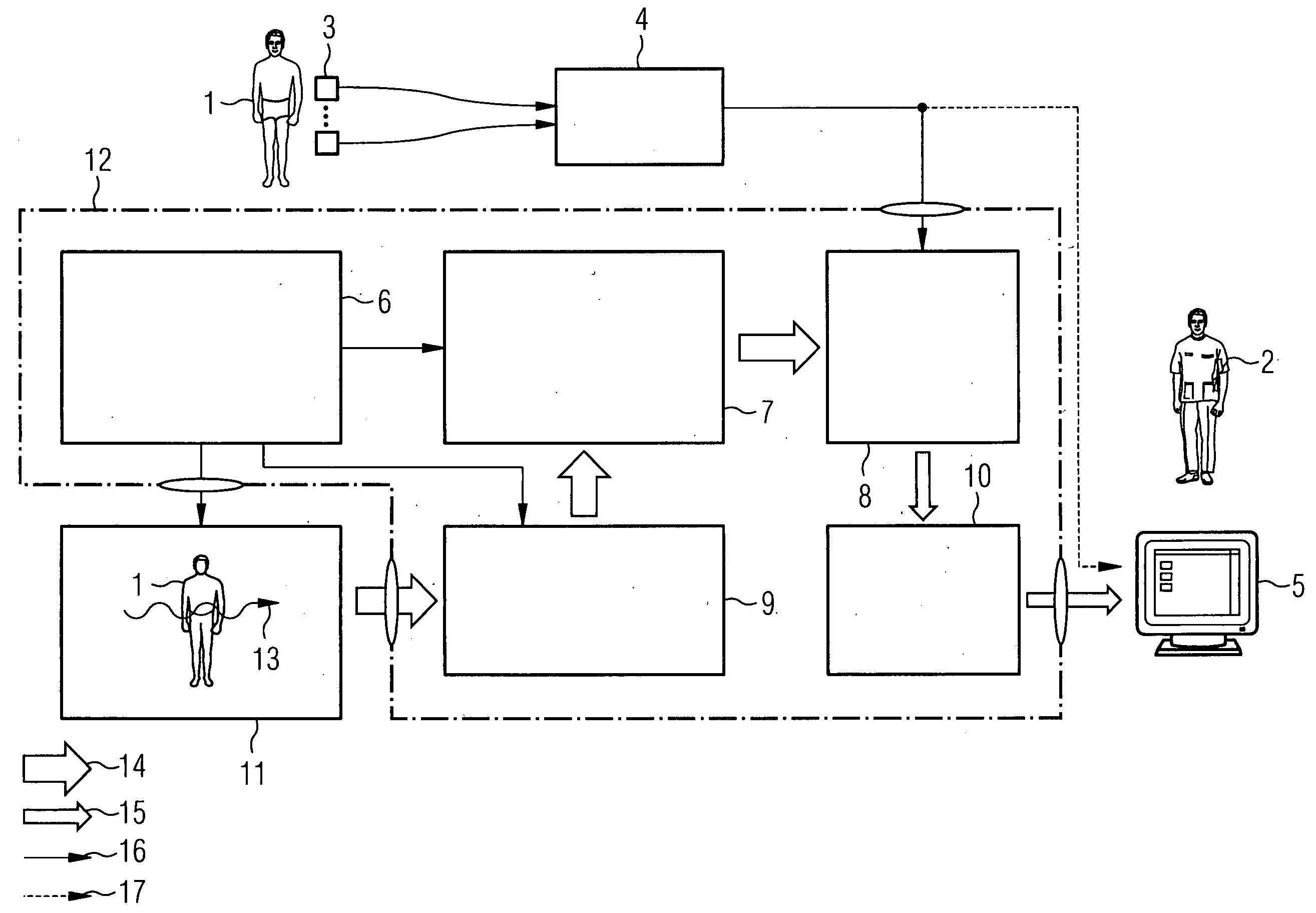
[0034]FIG. 1 shows a flowchart of the inventive method. The object of the inventive method is to undertake a type of fine registration at the actual object of interest of a series of images and to use the (balloon) marker registration only as a type of coarse pre-registration. The complete registration process therefore breaks down into a number of steps:

[0035]In a first step S1 the series of images is first recorded. This is done with any given medical Imaging modality which is suitable for creating informative images of the object of interest (e.g. stent 18) with the balloon markers A,B. The imaging modality 11 might for example be projection radiography, CT, MRT, US etc.

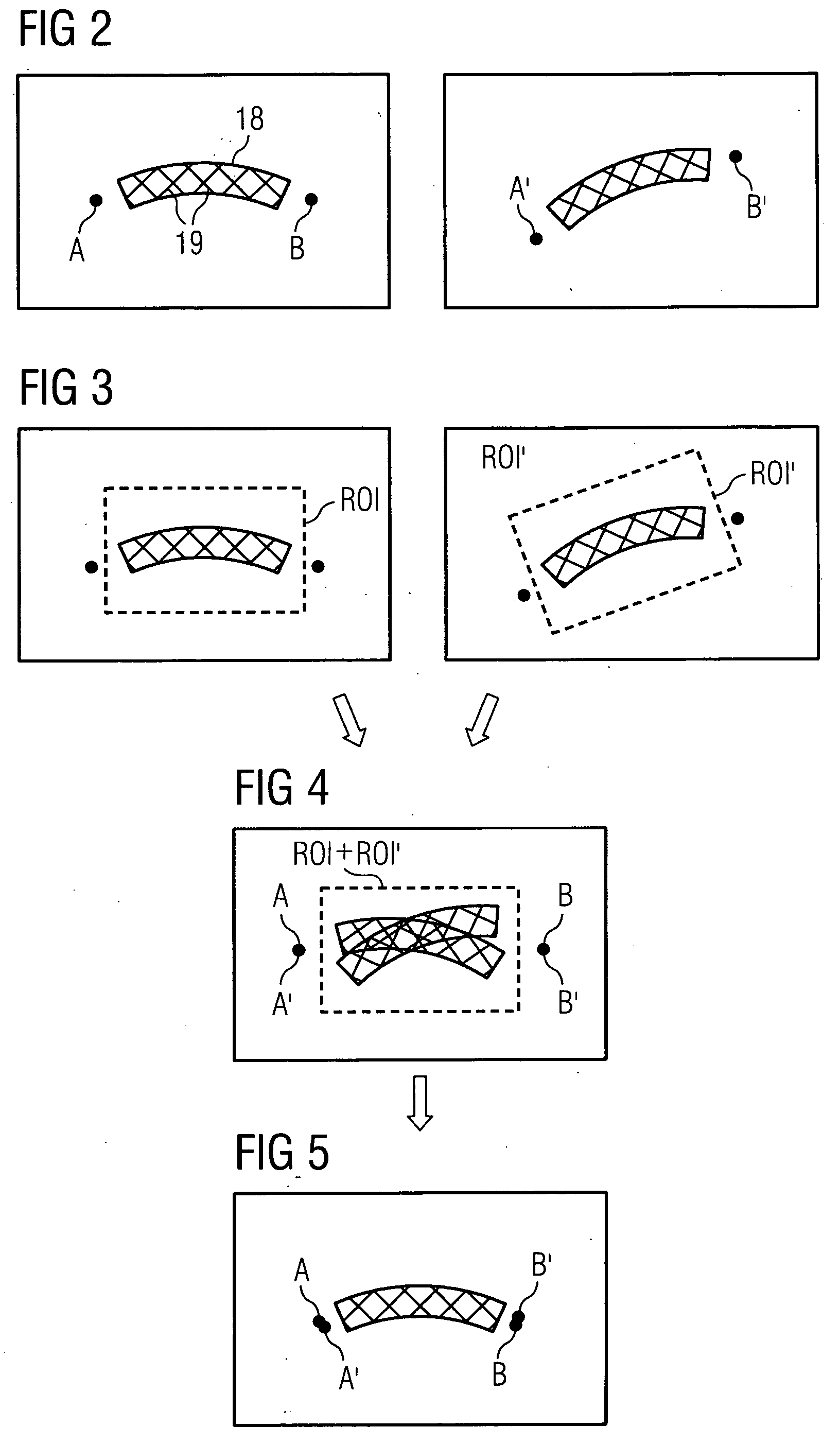
[0036]In a second step S2 an ROI (Region Of interest) is created in each image of the series of images and this is region is created (e.g. manually) in the form of a rectangle lying between the markers A, B of the balloon which entirely surrounds or entirely contains the object to be registered.

[0037]It can now be...


PUM


Abstract

The invention relates to a method for high-resolution display of vessel implants in angiographic images, featuring the steps: recording at least two images of an object-catheter combination including catheter markers with a medical imaging method; detection of an region of interest created in the form of an area between the balloon markers which completely contains the object to be registered for each recorded image; coarse registration of the region of interest images by registration of the respective pairs of balloon markers of all recorded images; fine registration of the region of interest image content/of the region of interest image by registration of the region of interest content; and arithmetic averaging across the fine-registered images.

Description

CROSS REFERENCE TO RELATED APPLICATIONS[0001]This application claims priority of German application No. 10 2007 019 328.0 filed Apr. 24, 2007, which is incorporated by reference herein in its entirety.FIELD OF THE INVENTION[0002]The present invention relates in general to an angiographic imaging method such as is used in medicine in the area of vascular surgery for example, more precisely in diagnostic and interventional cardiology. In such cases the present invention relates especially to a method which allows evaluation of the status during or after the placement of filigree vessel implants (such as stents for example) under the influence of different body movements.BACKGROUND OF THE INVENTION[0003]In modern (heart) vessel surgery (Percutaneous Transluminal (Coronary) Angioplasty PT(C)A expanding plastics—so-called stents—are inserted) to an increasing extent intravascularly into stenotized vessel sections. This involves implants usually in the form of thin wire meshes (wire diame...

Claims


Application Information

Patent Timeline
Patent Type & Authority Applications(United States)
IPC IPC(8): A61B5/00
CPCA61B5/7285A61B6/12A61B6/481A61B6/504A61B6/541A61B8/543G06T5/50G06T7/0028G06T2207/10116G06T2207/20016G06T2207/20132G06T2207/20216G06T2207/30052G06T2207/30101G06T2207/30204G06T7/33
Inventor LENDL, MARKUS
Owner SIEMENS HEALTHCARE GMBH
Why Eureka
Try Eureka