Methods and systems for attenuation correction in medical imaging

a technology of attenuation correction and medical imaging, applied in the field of medical imaging systems, can solve the problems of indeterminate or incorrect diagnosis of nodules, blurred nodule activity in pet scans, and affecting diagnosis, and image quality

Active Publication Date: 2008-11-06
GENERAL ELECTRIC CO
View PDF11 Cites 26 Cited by
  • Summary
  • Abstract
  • Description
  • Claims
  • Application Information

AI Technical Summary

Benefits of technology

[0004]A method for positron emission tomography / computed tomography (PET / CT) is provided. The method includes scanning an object / patient to acquire a stream of cine CT data and storing the data. The method further includes selecting a value for each pixel that represents the most dense measurement for the pixel (e.g. a cine (temporal) intensity maximum (CIM)) for each pixel throughout a cine CT acquisition. The method includes creating a single CT attenuation correction image composed of said CIM pixels. In another embodiment, the method also provides for the attenuation correction image to be reconstructed utilizing a weighted combination of a cine average CTAC and a CIM CTAC. Thus, undesirable motion artifacts can be removed from positron emission tomography (PET) images by utilizing the CT attenuation correction image.

Problems solved by technology

However, due to the comparatively lower resolution of PET images as compared to CT images, and due to the effects of respiratory or patient motion during a PET scan, nodule activity can be blurred in the PET scan.
Consequently, it can be difficult to quantify the nodule activity with a PET scan alone, which may result in an indeterminate or incorrect outcome of the diagnosis of the nodule.
Such image quality may affect diagnosis.
Misalignment of the CT attenuation map and PET emission image due to respiratory motion may cause errors in the attenuation correction (AC) factors and may produce artifacts in the final reconstructed AC PET image.
Thus, in the case of cardiac PET, attenuation artifacts may result in artificial visual depressions in the myocardial wall that may be incorrectly interpreted as perfusion defects by utilizing cine CT attenuation correction (CTAC) data.

Method used


Image

  • Methods and systems for attenuation correction in medical imaging
  • Methods and systems for attenuation correction in medical imaging
  • Methods and systems for attenuation correction in medical imaging

Examples


Embodiment Construction

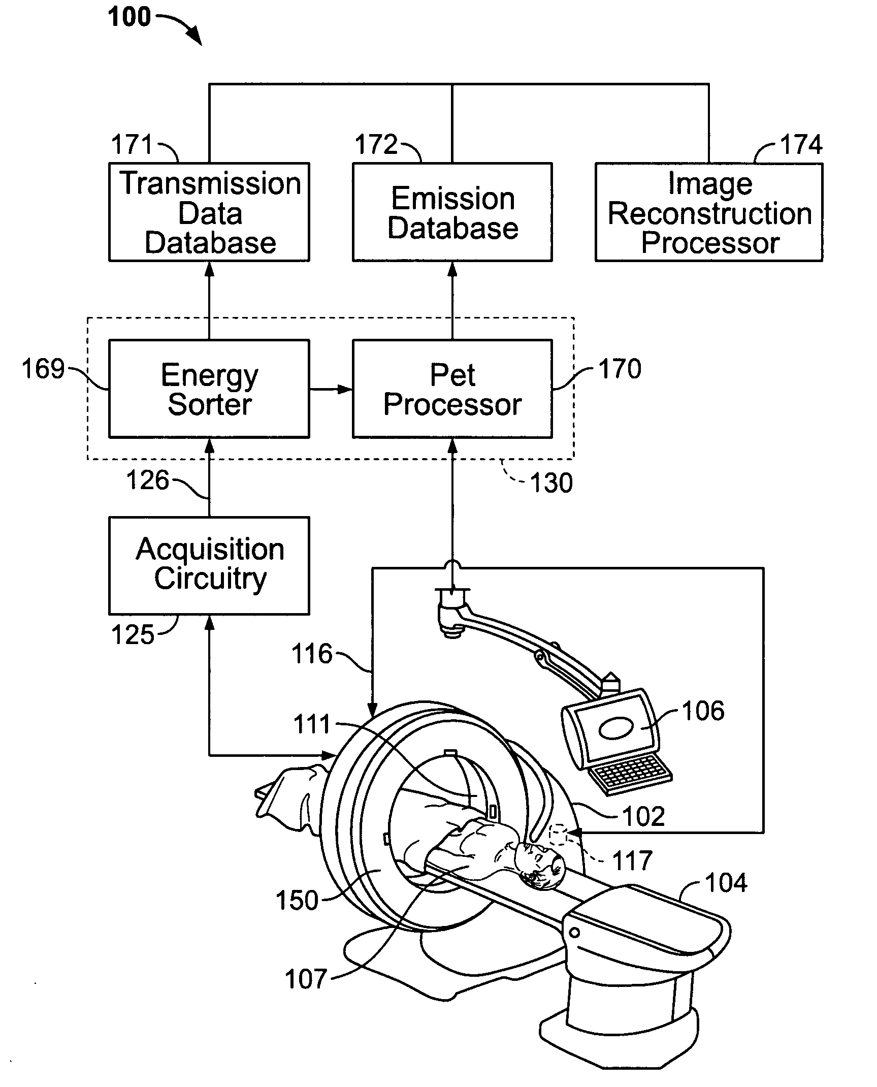
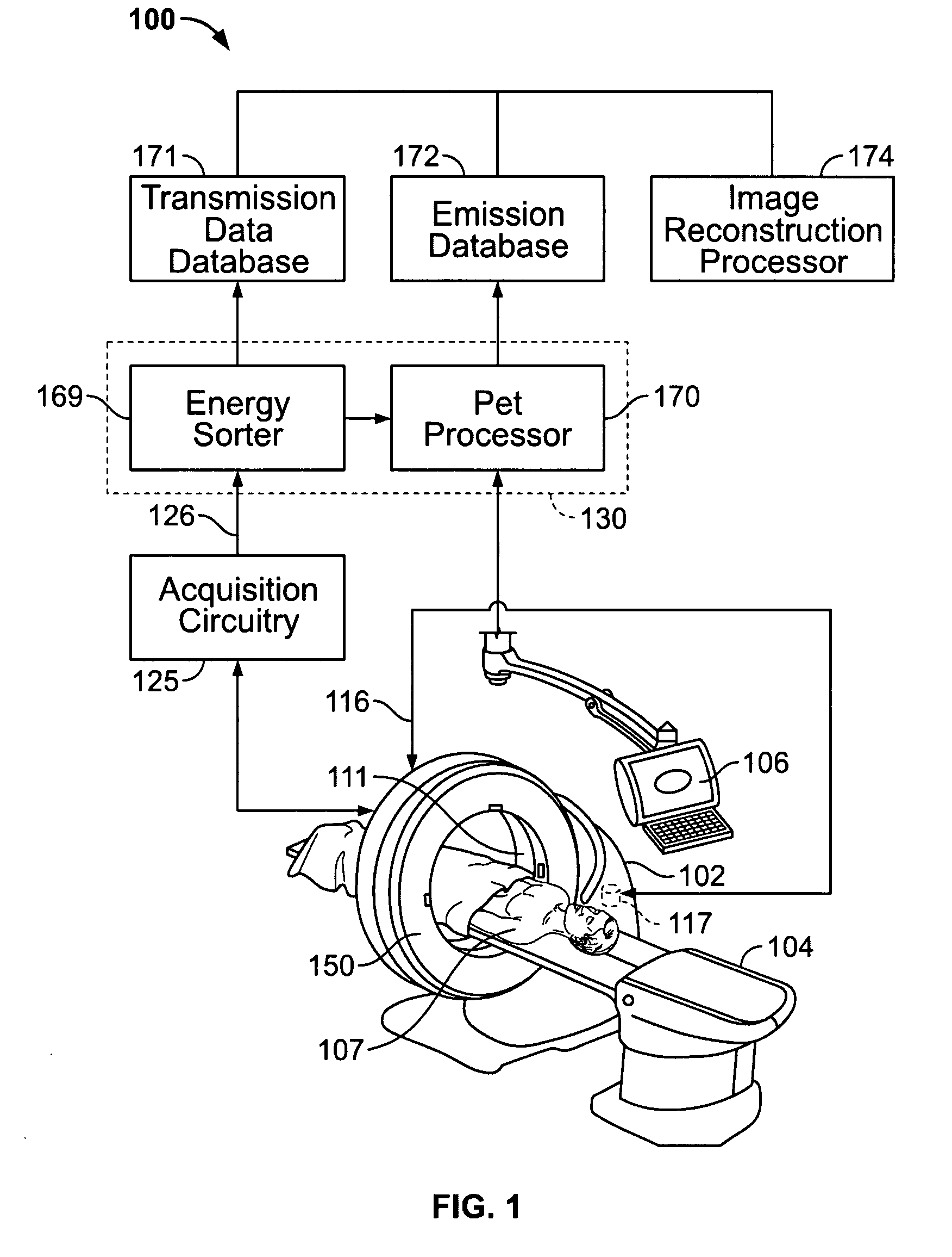
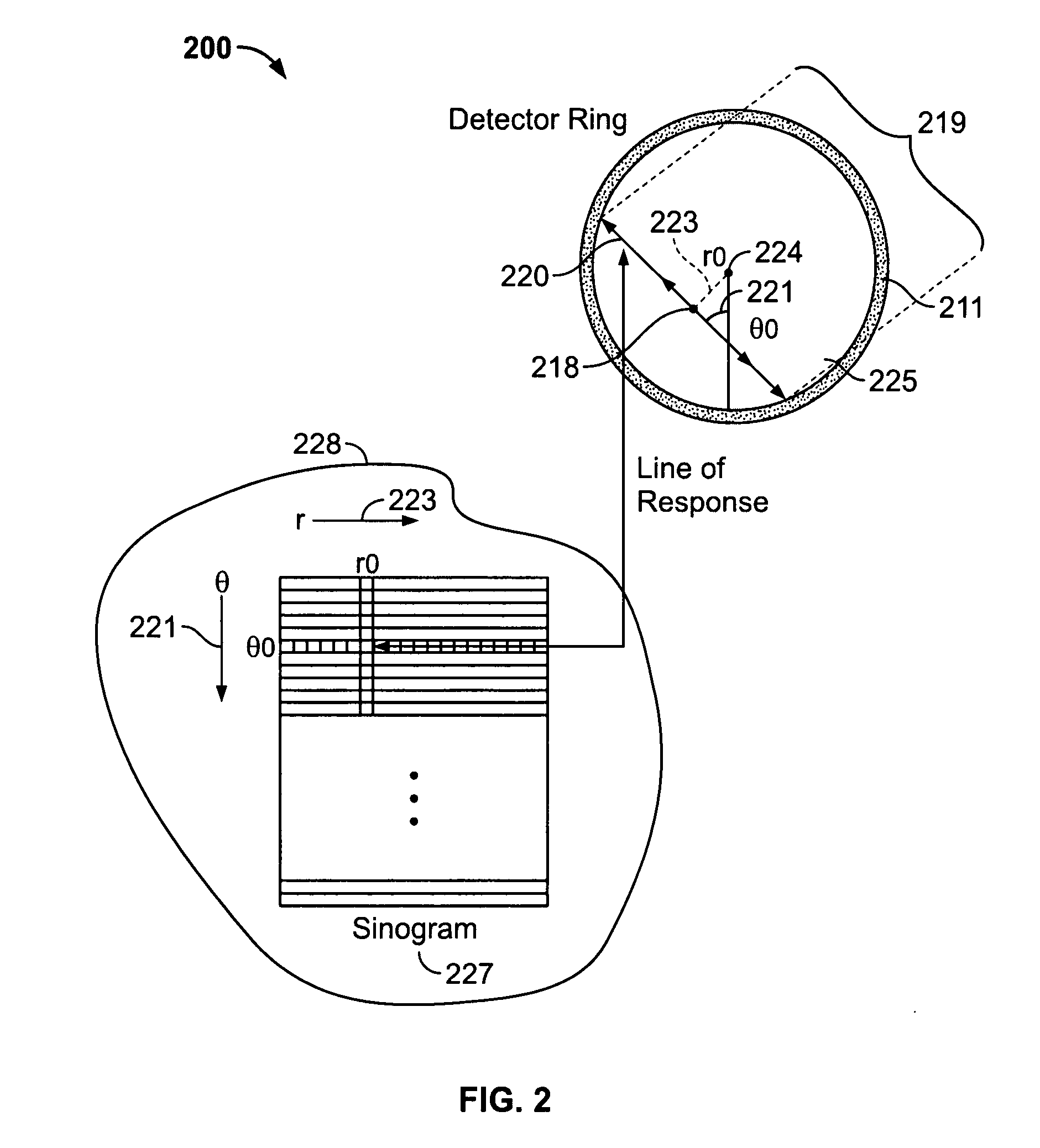
[0011]In the following detailed description, reference is made to the accompanying drawings which form a part hereof, and in which is shown by way of illustration specific embodiments in which the present invention may be practiced. These embodiments, which are also referred to herein as “examples,” are described in sufficient detail to enable those skilled in the art to practice the invention, and it is to be understood that the embodiments may be combined, or that other embodiments may be utilized and that structural, logical and electrical changes may be made without departing from the scope of the various embodiments of the present invention. The following detailed description is, therefore, not to be taken in a limiting sense, and the scope of the present invention is defined by the appended claims and their equivalents.

[0012]In this document, the terms “a” or “an” are used, to include one or more than one. In this document, the term “or” is used to refer to a nonexclusive or, ...


PUM


Abstract

Methods and systems for imaging a patient are provided. The method includes scanning a patient and acquiring a plurality of frames of cine computed tomography (CT) images during one complete respiratory cycle. In one embodiment, a method is provided that includes selecting a value for each pixel that represents the maximum density measurement for the pixel throughout the cine acquisition. In one embodiment, an attenuation correction image of a volume of interest is constructed by weighting a combination of the maximum pixel intensity value and an average pixel intensity value. Undesirable motion artifacts can be removed from positron emission tomography (PET) images by utilizing the CT attenuation correction image.

Description

BACKGROUND OF THE INVENTION[0001]This invention relates generally to medical imaging systems, and more particularly to attenuation correction for medical imaging.[0002]A nodule found during a CT scan often requires a patient to return many months later and obtain another CT scan to determine malignancy based on a nodule doubling time. PET scans may be helpful in diagnosis due to increased metabolic activity in the region of the nodule. However, due to the comparatively lower resolution of PET images as compared to CT images, and due to the effects of respiratory or patient motion during a PET scan, nodule activity can be blurred in the PET scan. Consequently, it can be difficult to quantify the nodule activity with a PET scan alone, which may result in an indeterminate or incorrect outcome of the diagnosis of the nodule.[0003]The image quality of at least some known PET and CT is highly affected by physiological patient moving. Such image quality may affect diagnosis. Lung nodules, ...

Claims


Application Information

Patent Timeline
Patent Type & Authority Applications(United States)
IPC IPC(8): G06K9/00
CPCA61B6/032A61B6/037A61B6/5235G06T11/005G06T2211/412
Inventor KOHLMYER, STEVEN GERALDALESSIO, ADAM MICHAELKINAHAN, PAUL EUGENEHURLEY, DENNIS PATRICK
Owner GENERAL ELECTRIC CO
Why Eureka
Try Eureka