Fourier-Domain Oct Ray-Tracing On The Eye

a fourier-domain oct ray and eye technology, applied in the field of measuring methods for ophthalmology, can solve problems such as motion artifacts and motion artifacts, and achieve the effect of fast measurement of reference points

Active Publication Date: 2008-11-20
CARL ZEISS MEDITEC AG
View PDF8 Cites 55 Cited by
  • Summary
  • Abstract
  • Description
  • Claims
  • Application Information

AI Technical Summary

Benefits of technology

[0020]Therefore, the primary object of the present invention is a very fast measurement of reference points for the three-dimensional dep

Problems solved by technology

This is time-consuming and can lead to motion artifacts.
However, this displacement, the required additional exposures and t

Method used


Image

  • Fourier-Domain Oct Ray-Tracing On The Eye
  • Fourier-Domain Oct Ray-Tracing On The Eye
  • Fourier-Domain Oct Ray-Tracing On The Eye

Examples


Embodiment Construction

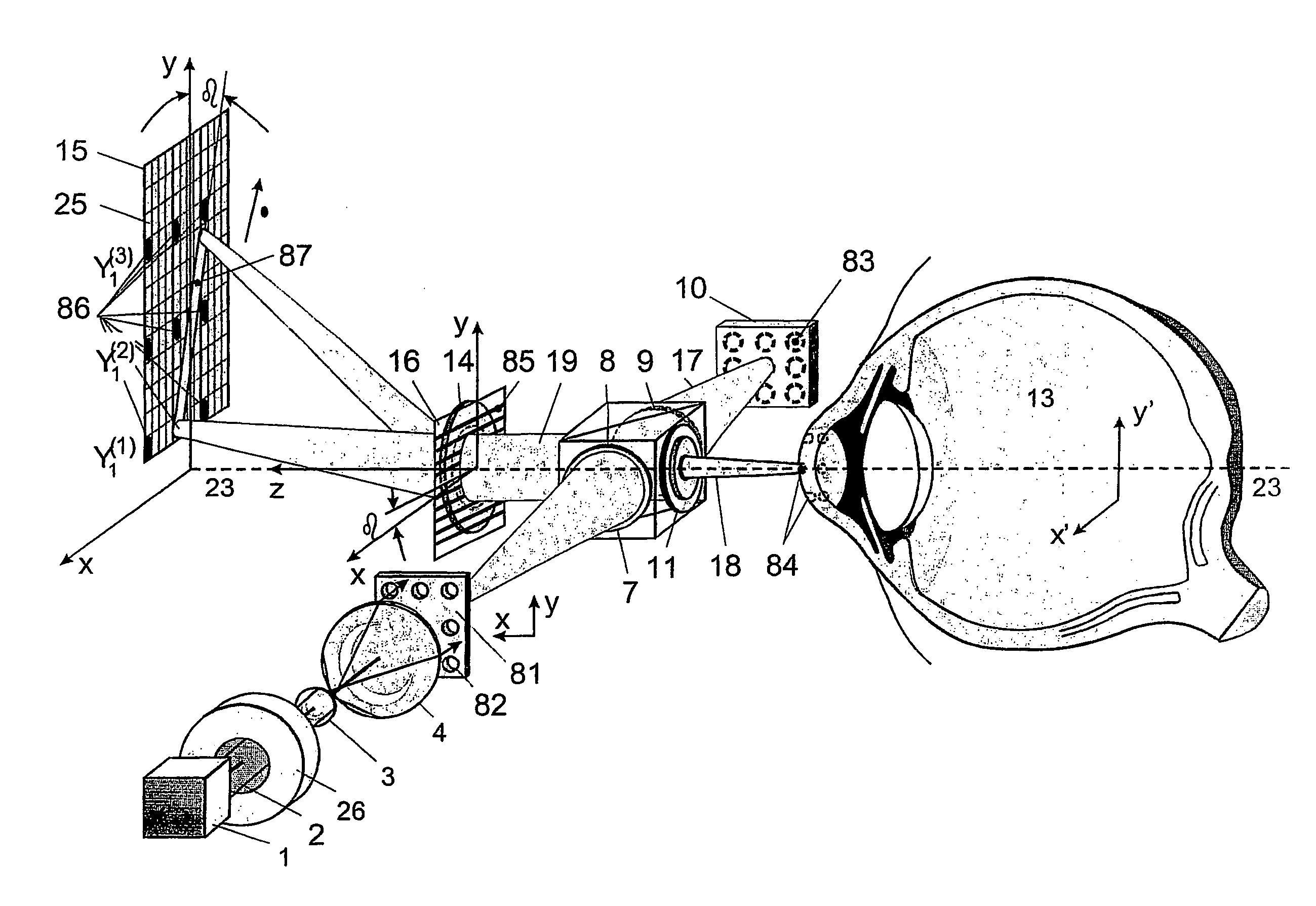
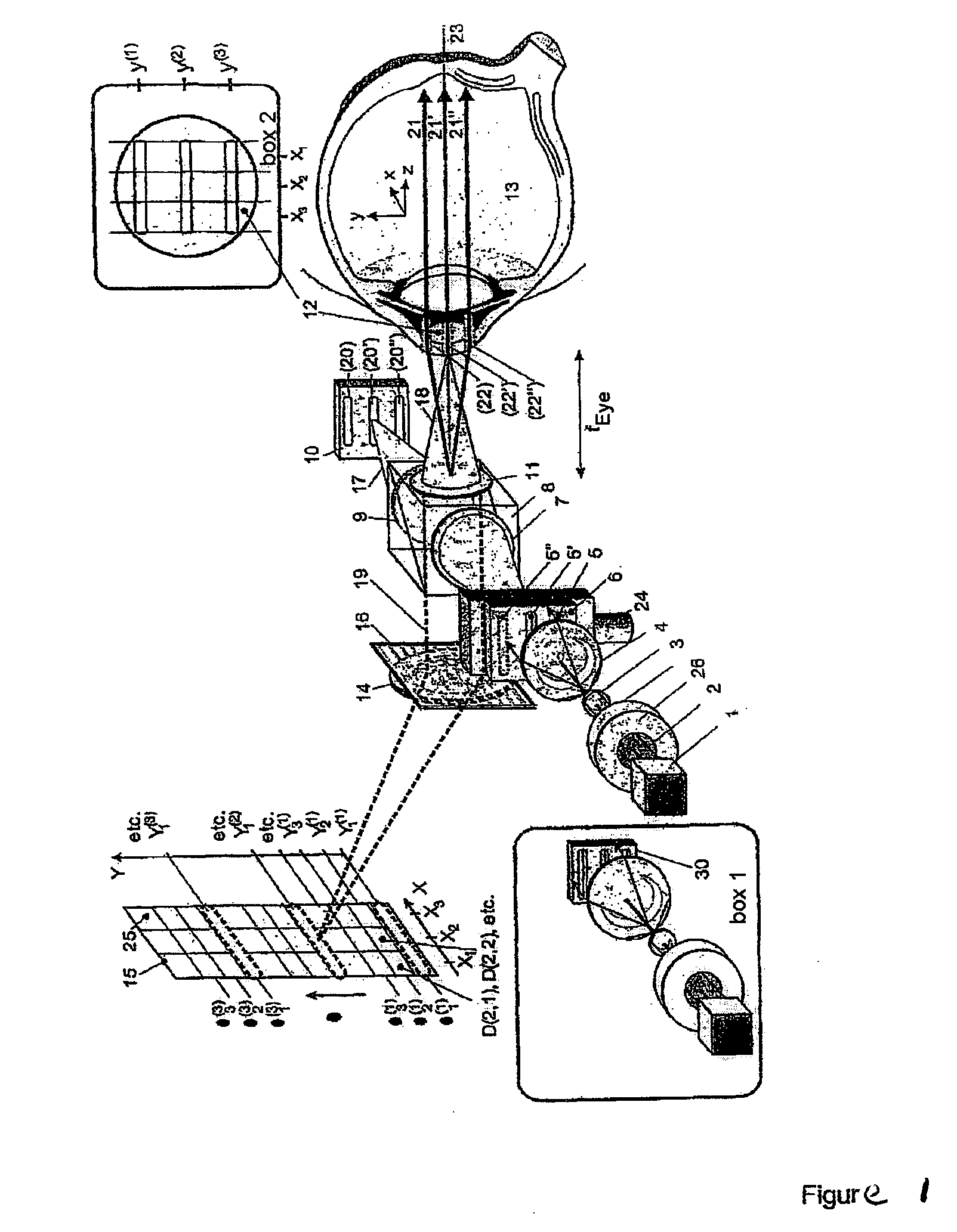
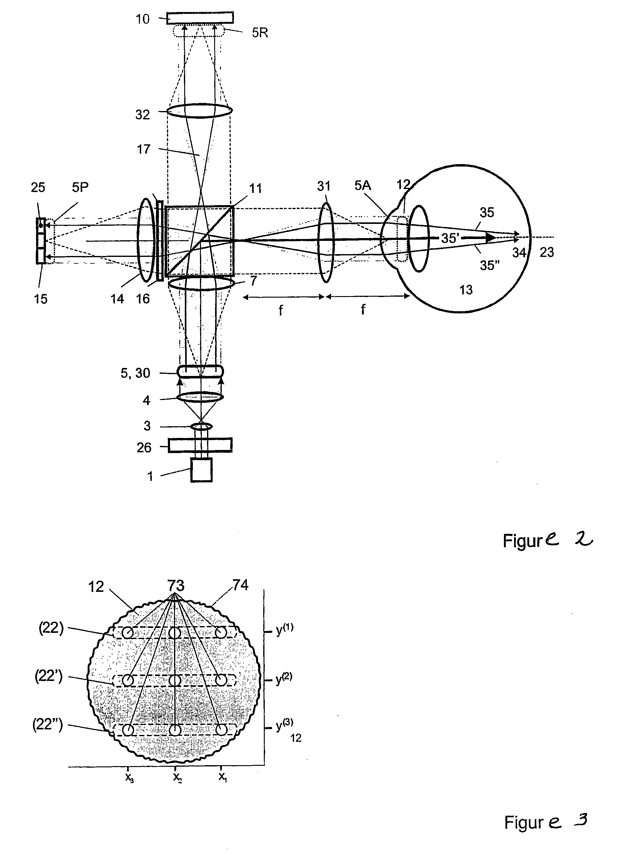
[0034]FIG. 1 shows a short-coherence interferometer according to the invention for ray tracing with divergent illumination of the eye. In this case, the light beam 2 which is emitted by the short-coherence illumination source 1 and passes through the shutter 26 illuminates, via optics 3 and 4, a light slit opening 5 which is moved in y-direction by a drive unit 24. The light slit diaphragm 6 can also have a series of openings instead of a slit. The light slit of the light slit diaphragm 6 is moved consecutively into positions 5, 5′ and 5″. The optics 7 image the light slit opening on the reference mirror 10 through the beamsplitter 8 by optics 9 on one side and on the pupil 12 of the eye 13 via the splitter surface of the beamsplitter 8 by optics 11 on the other side. The light slit images 20, 20′ and 20″ corresponding to the different positions 5, 5′ and 5″ of the light slit diaphragm 6 on the reference mirror 10 and 22, 22′ and 22″ in the pupil 12 of the eye 13 are imaged by optic...


PUM


Abstract

The present invention is directed to an ophthalmologic measurement method which can depict three-dimensional structures of the interfaces of an eye by short-coherence interferometry based on reference points. For this purpose, the pupil is illuminated at a plurality of points by a short-coherence illumination source. The measurement beam reflected at these points by the interfaces and surfaces of the eye is superimposed with a reference beam. The measurement data which are generated in this way are spectrally split by a diffraction grating, imaged on a two-dimensional detector array and conveyed to a control unit which determines a three-dimensional structure of all intraocular interfaces and surfaces of the eye. In the suggested Fourier domain OCT method, the depiction of three-dimensional structures is preferably carried out by spline surfaces or polygon surfaces. In doing so, it is possible to determine the depth positions of the measurement beams in many points of the pupil with a single recording of the array camera in that the pupils are illuminated by a diaphragm grid and the reference mirror contains a periodic phase grid.

Description

CROSS-REFERENCE TO RELATED APPLICATIONS[0001]This application claims priority of International Application No. PCT / EP2005 / 008090, filed Jul. 26, 2005 and German Application No. 10 2004 037 479.1, filed Aug. 3, 2004, the complete disclosures of which are hereby incorporated by reference.BACKGROUND OF THE INVENTION[0002]a) Field of the Invention[0003]The present invention is directed to a measuring method for ophthalmology which uses short-coherence interferometry to measure the positions of reference points on the basis of which the three-dimensional structure of all refracting and reflecting intraocular interfaces and surfaces can be depicted, for example, by means of spline surfaces or polygon surfaces.[0004]b) Description of the Related Art[0005]For this purpose, measurement beams are radiated into the eye simultaneously through a number of pupil points, and the depth positions at which these measurement beams pierce or are reflected at intraocular interfaces and surfaces are dete...

Claims


Application Information

Patent Timeline
IPC IPC(8): A61B3/107
CPCA61B3/102A61B3/103
Inventor FERCHER, ADOLF FRIEDRICH
Owner CARL ZEISS MEDITEC AG
Why Eureka
Try Eureka