Identifying Ribs in Lung X-Rays

Inactive Publication Date: 2008-12-25
SIEMENS COMP AIDED DIAGNOSIS +1
View PDF10 Cites 36 Cited by
  • Summary
  • Abstract
  • Description
  • Claims
  • Application Information

AI Technical Summary

Benefits of technology

[0024]iv. categorizing and elimi

Problems solved by technology

Although supposed to keep still and hold their breadth, keeping lungs inflated, patients may move or inhale or exhale during the exposure to x-rays, blurring the resultant radiography image somewhat.
Since x-ray radiation may itself cause malignancies, radiologists are wary of repeatedly exposing patients to x-rays, particularly with pregnant women, the young, the elderly and/or the ill.
With some systems, such as those using traditional film, it may be impossible to repeat the test once a problem is determined, as the patient is no longer ava

Method used


Image

  • Identifying Ribs in Lung X-Rays
  • Identifying Ribs in Lung X-Rays
  • Identifying Ribs in Lung X-Rays

Examples


Embodiment Construction

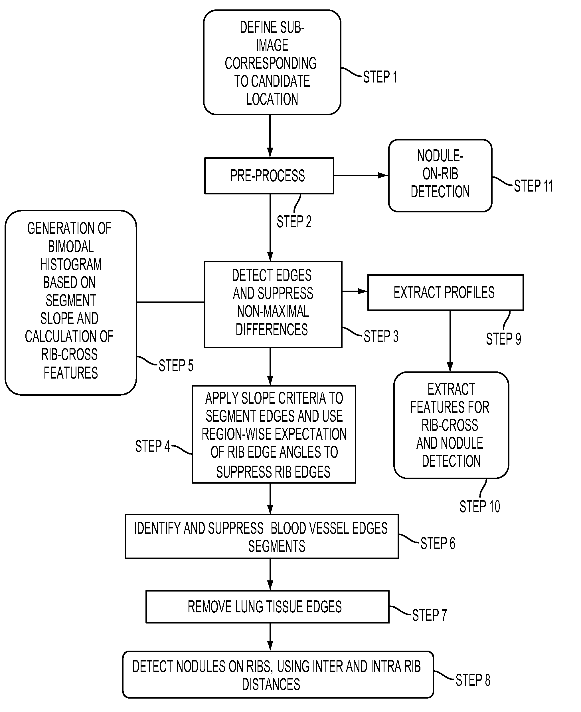
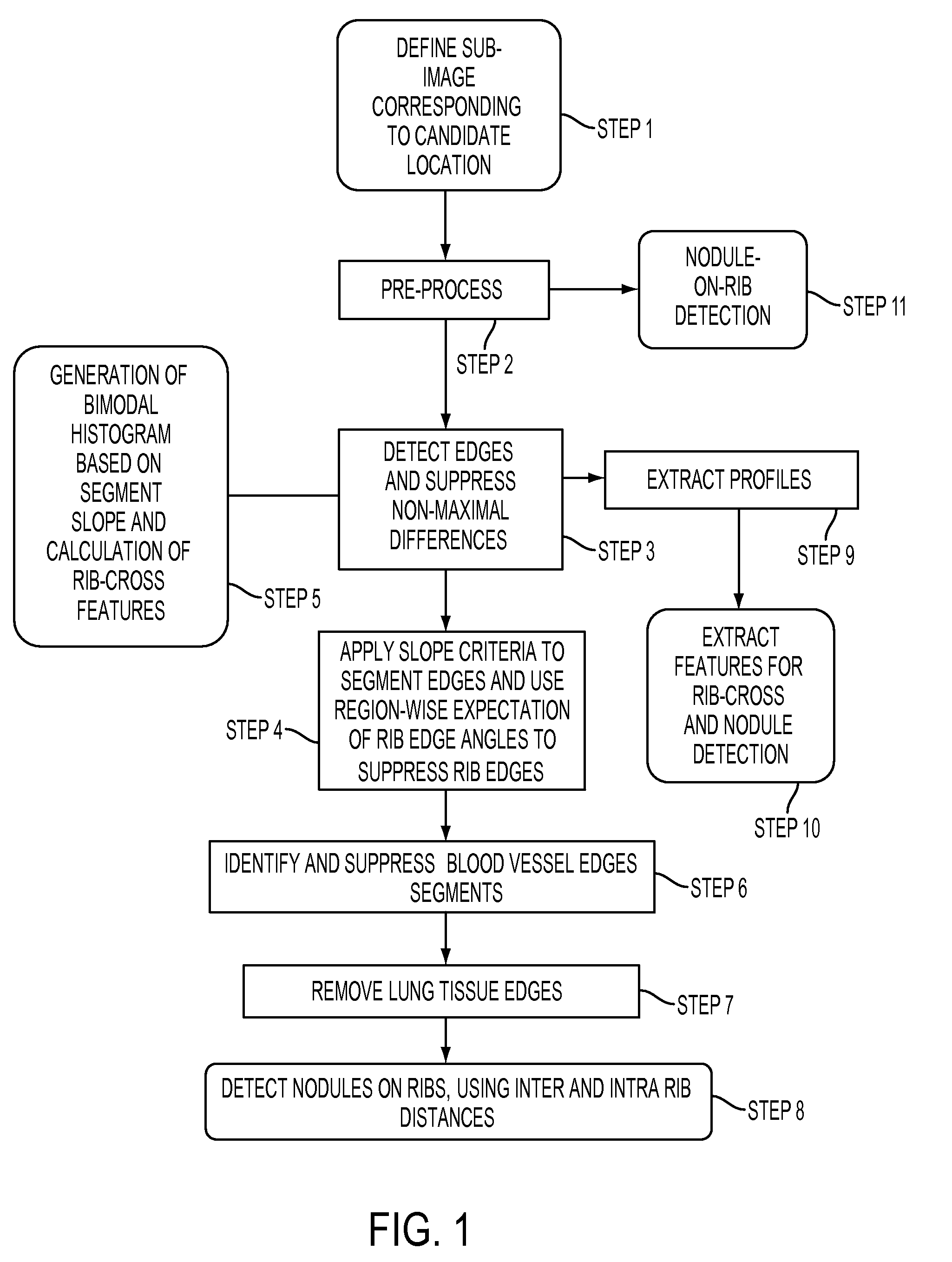
[0075]The shadows of blood vessels and ribs in posterior anterior x-ray radiography images of the chest obscure nodules, making them difficult to identify.

[0076]Embodiments of the present invention are directed to detecting, identifying and correctly characterizing physiological features, particularly ribs and blood vessels which show up in x-ray radiographs, obscuring nodules and other irregularities of interest. Applications include training classifiers, improved CAD systems and faster more accurate diagnostics.

[0077]An edge detection scheme is employed. When looking for nodules and the like, by attributing edges appearing in posterior anterior x-ray images to ribs, blood vessels or edges of the lungs themselves, these may be discounted. Non-discounted edges are more likely to be nodules requiring further consideration. In this manner, the ratio of nodules to false positives is maximized and the effectiveness of CAD image analysis is improved.

[0078]Correct identification of candid...


PUM


Abstract

A method of detecting lung nodules in an anterior posterior x-ray radiograph comprising the steps of: generating candidate regions in image showing changes in contrast above a threshold level, and eliminating false positives by eliminating edges assignable to organs by: identifying edges; categorizing and eliminating rib edges; categorizing and eliminating lung tissue edges, and categorizing and eliminating blood vessels.

Description

CROSS REFERENCE TO RELATED APPLICATION[0001]The present application claims priority rights from U.S. Provisional Application No. 60 / 941,826, filed Jun. 4, 2007; U.S. Provisional Application No. 60 / 941,801, filed Jun. 4, 2007; and U.S. Provisional Application No. 60 / 941,811, filed Jun. 4, 2007.FIELD OF THE INVENTION[0002]The present invention relates to computer aided diagnosis (CAD), and particularly to imaging the thoracic cavity for lung examination purposes.BACKGROUND TO THE INVENTION[0003]The chest x-ray is the most commonly performed diagnostic x-ray examination. The heart, lungs, airway, blood vessels and the bones of the spine and chest are imaged in a painless medical test that helps in the diagnosis of medical conditions.[0004]The chest x-ray is typically the first imaging test used to help diagnose causes of symptoms such as shortness of breath, fever, a bad or persistent cough, chest pain or injury. Its application helps in diagnosing and monitoring treatment for medical ...

Claims


Application Information

Patent Timeline
IPC IPC(8): G06K9/00
CPCG06T7/0012G06T2207/10116G06T2207/30061G06T2207/30064G06T7/13
Inventor ACHARYYA, MAUSUMICHAKRAVARTY, SUMITSIDDU, DINESH MYSORERATNER, ELIAHUMANEVITCH, ALEXANDRASTOECKEL, JONATHAN
Owner SIEMENS COMP AIDED DIAGNOSIS
Why Eureka
Try Eureka