X-ray imaging apparatus

a technology of x-ray imaging and x-ray, which is applied in the field of x-ray imaging apparatus, can solve the problems of ctha as soft tissue imaging, ct is inferior to ct in density resolution, and requires a large amount of contrast medium

Active Publication Date: 2009-01-22
TOSHIBA MEDICAL SYST CORP
View PDF1 Cites 8 Cited by
  • Summary
  • Abstract
  • Description
  • Claims
  • Application Information

AI Technical Summary

Benefits of technology

[0009]It is an object of the present invention to provide an X-ray imaging apparatus which can generate two types of three-dimensional images by one rotation radiography.

Problems solved by technology

An IVR-CT is very expensive, and hence only some large hospitals can purchase it.
CTHA as soft tissue imaging is, however, inferior to CT in density resolution, and hence requires a larger amount of contrast medium.
It is therefore feared that an increasing amount of contrast medium will increase the burden on a patient.

Method used


Image

  • X-ray imaging apparatus
  • X-ray imaging apparatus
  • X-ray imaging apparatus

Examples


Embodiment Construction

[0017]An X-ray imaging apparatus according to a preferred embodiment of the present invention will be described below with reference to the views of the accompanying drawing.

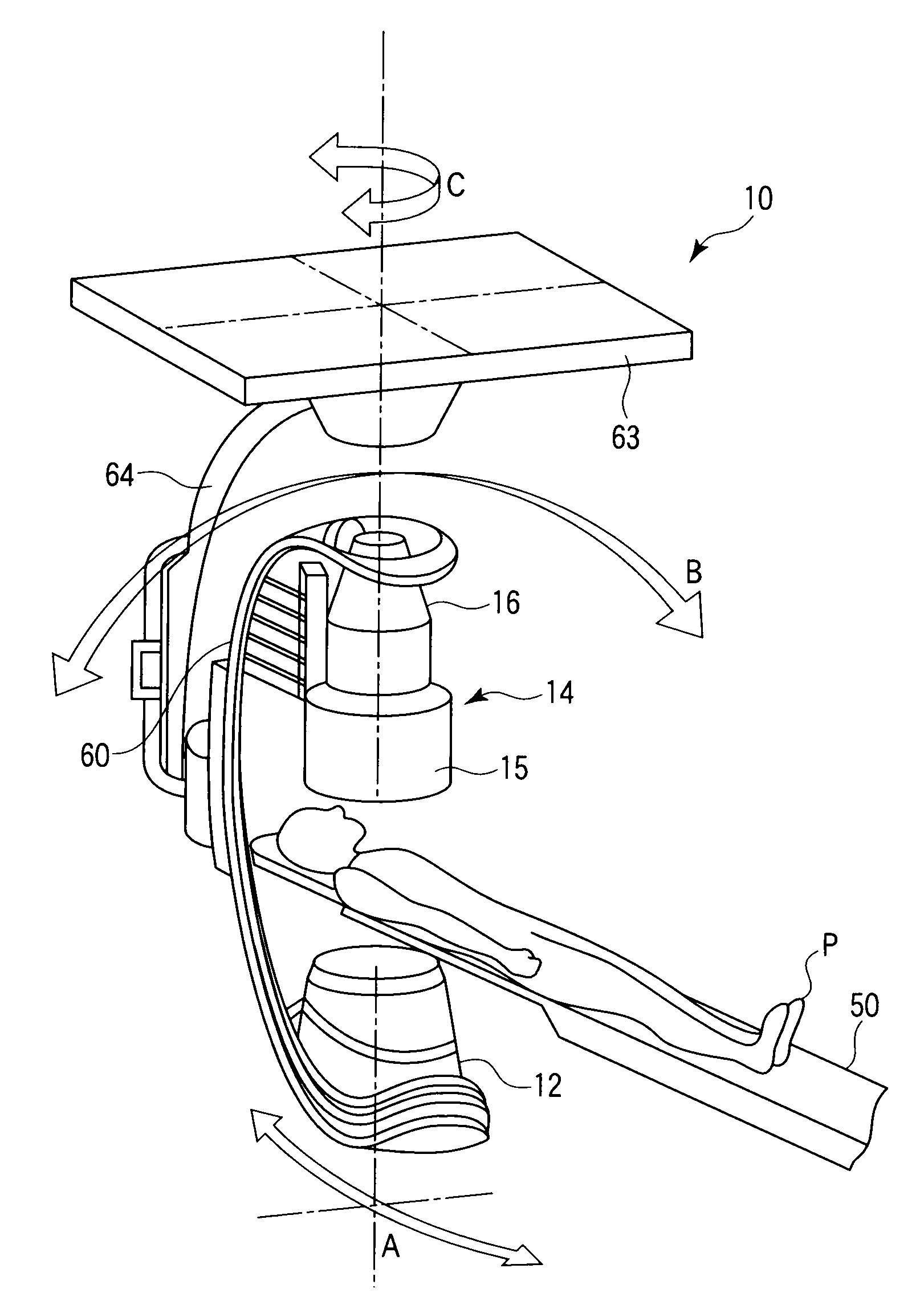
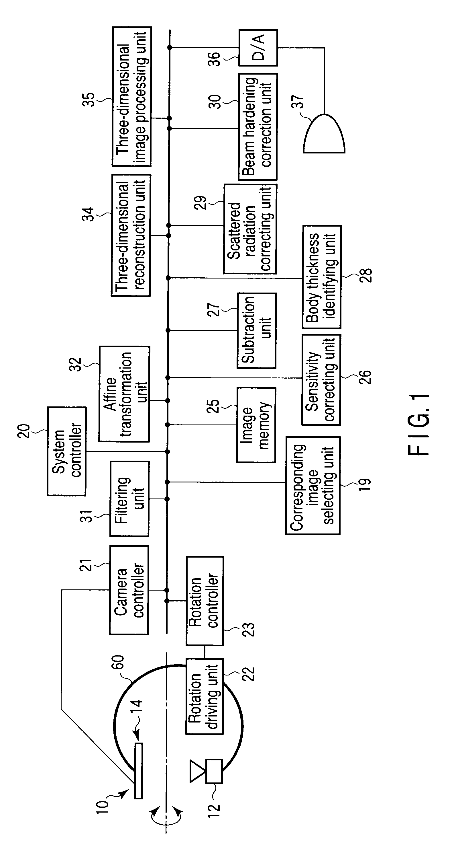
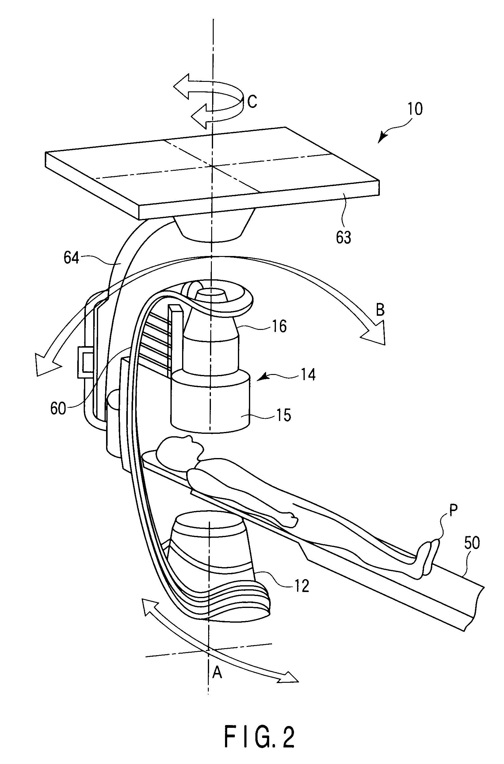
[0018]As shown in FIG. 1, the X-ray imaging apparatus includes an X-ray imaging mechanism 10. As shown in FIG. 2, the X-ray imaging mechanism 10 includes an X-ray tube 12 and an X-ray detector 14. The X-ray detector 14 comprises an image intensifier 15 and a TV camera 16. Alternatively, the X-ray detector 14 comprises a flat panel detector (FPD) having semiconductor detection elements arrayed in a matrix form. The X-ray tube 12 and the X-ray detector 14 are mounted on a C-arm 60 so as to face each other. A subject P on a top 50 of a bed is placed between the X-ray tube 12 and the X-ray detector 14. The C-arm 60 is supported by a column 64 suspended from a ceiling base 63 or by a floor-type stand. The C-arm 60 is rotatable with respect to three orthogonal axes A, B, and C. A rotation driving unit 22 is housed in ...


PUM


Abstract

An X-ray imaging apparatus includes a substantially C-arm, a support mechanism which rotatably supports substantially the C-arm, a rotation driving unit which drives rotation of substantially the C-arm, an X-ray tube mounted on substantially the C-arm, an X-ray detector mounted on substantially the C-arm, and a control unit which controls at least one of the rotation driving unit and an imaging control unit to make intervals between a plurality of contrast-enhanced images shorter than intervals between a plurality of mask images by changing a rotational speed of substantially the C-arm before and after injection of a contrast medium.

Description

CROSS-REFERENCE TO RELATED APPLICATIONS[0001]This application is based upon and claims the benefit of priority from prior Japanese Patent Application No. 2007-187426, filed Jul. 18, 2007, the entire contents of which are incorporated herein by reference.BACKGROUND OF THE INVENTION[0002]1. Field of the Invention[0003]The present invention relates to an X-ray imaging apparatus which generates a three-dimensional image of a blood vessel from images before and after the injection of a contrast medium.[0004]2. Description of the Related Art[0005]There is an examination technique called CTHA (CT during hepatic arteriography) for diagnosing a liver tumor. This is a method of performing CT (Computed Tomography) of a hepatic artery while injecting a contrast medium into it. In general, however, this technique can be implemented by only an apparatus (called an IVR-CT) comprising both a CT apparatus and an angiography apparatus. An IVR-CT is very expensive, and hence only some large hospitals ...

Claims


Application Information

Patent Timeline
Patent Type & Authority Applications(United States)
IPC IPC(8): A61B6/00G06K9/00
CPCA61B5/4244A61B6/4441A61B6/504A61B6/466A61B6/481A61B6/4464Y10S378/901
Inventor OHISHI, SATORUHIROSE, MASASHI
Owner TOSHIBA MEDICAL SYST CORP
Why Eureka
Try Eureka