Deployment of polysaccharide markers

a polysaccharide and marker technology, applied in the field of polysaccharide markers, can solve the problems of inaccuracy, misdirected follow-up treatment to an undesired portion of the patient's tissue, and rapid hemostasis, and no long-term irritation or inflammation

Inactive Publication Date: 2009-01-29
SENORX
View PDF99 Cites 44 Cited by
  • Summary
  • Abstract
  • Description
  • Claims
  • Application Information

AI Technical Summary

Benefits of technology

[0016]The polysaccharide containing pellets will generally have a transverse dimension of about 0.02 to about 0.1 inch (0.5-2.5 mm), preferably about 0.035 to about 0.075 inch (0.9-1.9 mm), and typically about 0.056 inch (1.4 mm). The pellets will have a length of about 0.1 to about 0.4 inch (2.5-10 mm), preferably about 0.15 to about 0.35 inch (3.8-8.9 mm). The pellets provide sufficient column strength to facilitate introduction into and discharge thereof from the tubular delivery device. The markers are preferably arranged for delivery within the delivery tube with the marker having a radiopaque element between two adjacent marker members formed of polysaccharide. As presently contemplated, within the delivery tube there will be two polysaccharide marker members distal and two polysaccharide marker members proximal to the marker having a radiopaque element.
[0018]Upon being discharged into the intracorporeal target site, the plurality of polysaccharide containing markers quickly take up body fluid at the site, initiating the clotting process and providing hemostasis. The other marker member with a radiopaque marker element enables short term detection (at least three weeks, preferably at least four weeks but less than a year) by remote USI and preferably long term detection by remote mammographic imaging or MRI identification by the radiopaque element. Typically, the polysaccharide bodies dissolve in situ in about five seconds to about 2 minutes and enzymatically degrade in about 2-5 days.
[0023]The polysaccharide containing markers embodying features of the present invention provide several advantages. The polysaccharide marker bodies quickly dissolve in the body fluids at the site to provide to provide rapid hemostasis therein even with serious bleeding. Moreover, the materials rapidly degrade so there is no long term irritation or inflammation at the site.

Problems solved by technology

However, radiographically visible tissue features, originally observed in a mammogram, may be removed, altered or obscured by the biopsy procedure, and may heal or otherwise become altered following the biopsy.
This, in turn, can result in follow-up treatments being misdirected to an undesired portion of the patient's tissue.
Such a marker enables a follow-up procedure to be performed without the need for traditional radiographic mammography imaging which, as discussed above, can be subject to inaccuracies as a result of shifting of the location wire as well as being tedious and uncomfortable for the patient.

Method used


Image

  • Deployment of polysaccharide markers
  • Deployment of polysaccharide markers

Examples


Embodiment Construction

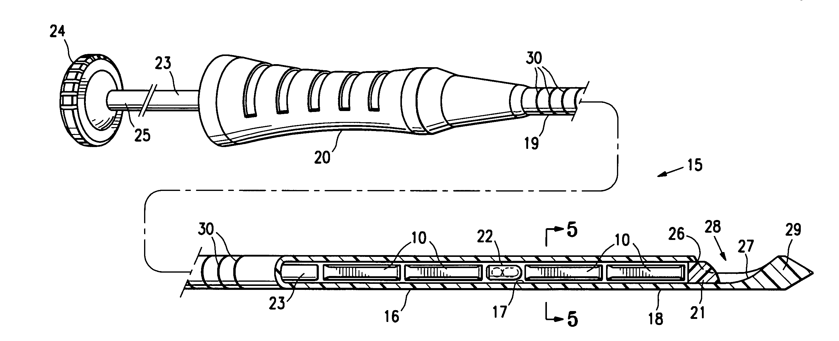
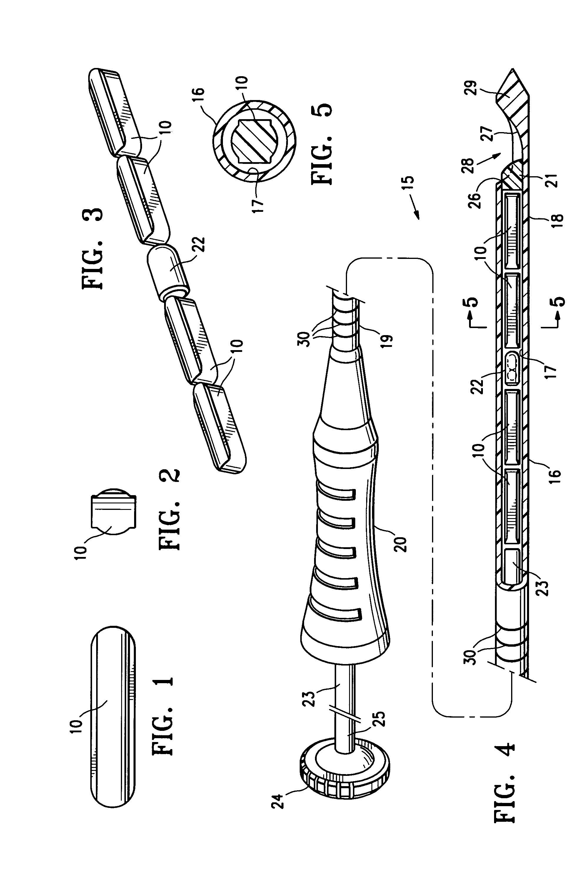
[0030]FIGS. 1 and 2 illustrate a press-formed marker member 10 embodying features of the invention which is formed of a mixture of polysaccharide powder and methylcellulose powder. The press-formed marker member 10 has sufficient polysaccharide powder so as to quickly form thrombus when coming into contact with blood at an intracorporeal site. Typically, the marker member 10 will have 65% (by wt.) polysaccharide and 35% (by wt.) methylcellulose. The marker member 10 is preferably formed by mixing polysaccharide powder (corn starch or potato starch) and methylcellulose powder in appropriate amounts, placing the mixed powder in a pellet die and subjecting the powder within the pellet die to a pressure of about 6 to 40 ksi, typically about 12 ksi.

[0031]One suitable polysaccharide material is U.S.P. Topical Starch. Alternatively, Hemaderm™, which is available from Medafor, Inc. located in Minneapolis, Minn., may also be used. This product is described at least in part in U.S. Pat. No. 6...


PUM


Abstract

The marker member delivery system described has a plurality of marker members disposed within a delivery tube or cannula which contain a suitable polysaccharide such as starch (e.g. corn starch or potato starch) and a binder such as methylcellulose. These marker members are preferably press-formed from powders. The system has preferably has at least one other marker member formed of bioabsorbable material with a radiopaque element attached to or incorporated therein. The marker with a radiopaque marker element is preferably disposed within the delivery tube between two marker members formed of polysaccharide and is preferably formed of a bioabsorbable material other than a polysaccharide.

Description

RELATED APPLICATIONS[0001]This application is related to and claims priority from provisional application Ser. No. 60 / 835,740, filed on Aug. 4, 2006, and copending application Ser. No. 11 / 881,264, filed on Jul. 26, 2007, both of which are incorporated herein by reference in their entirety.FIELD OF THE INVENTION[0002]The invention is generally directed to remotely detectable, intracorporeal markers and devices and methods for the delivery of such markers to a desired location within a patient's body.BACKGROUND OF THE INVENTION[0003]In diagnosing and treating certain medical conditions, it is often desirable to mark a suspicious body site for the subsequent taking of a biopsy specimen, for delivery of medicine, radiation, or other treatment, for the relocation of a site from which a biopsy specimen was taken, or at which some other procedure was performed. As is known, obtaining a tissue sample by biopsy and the subsequent examination are typically employed in the diagnosis of cancers...

Claims


Application Information

Patent Timeline
Patent Type & Authority Applications(United States)
IPC IPC(8): A61B6/00
CPCA61B19/54A61B2017/00004A61B2017/00831A61B2019/5487A61B2019/5408A61B2019/5425A61B2019/5462A61B2017/00898A61B90/39A61B2090/3908A61B2090/3925A61B2090/3962A61B2090/3987A61P7/04A61K9/0024
Inventor JONES, MICHAEL L.LUBOCK, PAUL
Owner SENORX
Why Eureka
Try Eureka