
Composition for the delivery of live cells and methods of use thereof

a technology for live cells and liquid systems, applied in drug compositions, skeletal/connective tissue cells, metabolic disorders, etc., can solve the problems of difficult control of liquid systems within a particular area, risk of infection, invasive and painful procedures, etc., and achieve the effect of improving the primary therapeutic effect and shortening the time period

Inactive Publication Date: 2009-02-12
COSTANTINO HENRY R +2
View PDF6 Cites 4 Cited by
  • Summary
  • Abstract
  • Description
  • Claims
  • Application Information

AI Technical Summary

Benefits of technology

The invention is a method and composition for administering live cells to a patient in need of treatment. The composition includes live cells and biocompatible, biodegradable polymer microparticles that can be injected into a treatment site or implantation site. The cells can be administered with or without a biologically active agent incorporated into the microparticles. The method and composition provide a means for eliciting a therapeutic effect in a patient without the need for open surgical intervention. The invention can be used for the treatment of cartilage deficiencies, defects, voids, and conformational discontinuities in a patient. The cells used in the invention can be live pancreatic islet cells, dopaminergic cells, or other suitable cells. The composition can also be used to generate new tissue growth or produce a biologically active molecule in a patient.

Problems solved by technology

However, implantation of the scaffolds requires surgical intervention which presents disadvantages, such as the risk of infection and the need for invasive and painful procedures.
However, controlled delivery and containment of a liquid system within a particular area is difficult, and the liquid can spread to areas other than the implant site prior to solidification.

Method used


Image

  • Composition for the delivery of live cells and methods of use thereof
  • Composition for the delivery of live cells and methods of use thereof
  • Composition for the delivery of live cells and methods of use thereof

Examples


example 1

Preparation of Microparticles

[0096]Microparticles were fabricated from PLG (50:50 lactide:glycolide, uncapped (—COOH), Mw˜10 kDa) using the General Process outlined above.

example 2

Cell Isolation-Chondrocytes

[0097]Bovine articular cartilage was harvested from the gelnohumeral and humeroulnar joints of neonatal calves and digested in 0.3% Type II collagenase at 37° C. for 12-16 hours overnight with shaking. Chondrocytes were passed through a 180 ÿm filter to remove large particulate material. Cells were washed 3 times with PBS and resuspended in complete medium (Ham's F12, 10% FBS, 0.3% carboxymethylcellulose, pen / strep / amphotericin B, and ascorbic acid).

example 3

Chondrocyte / Microparticle Adhesion Assay

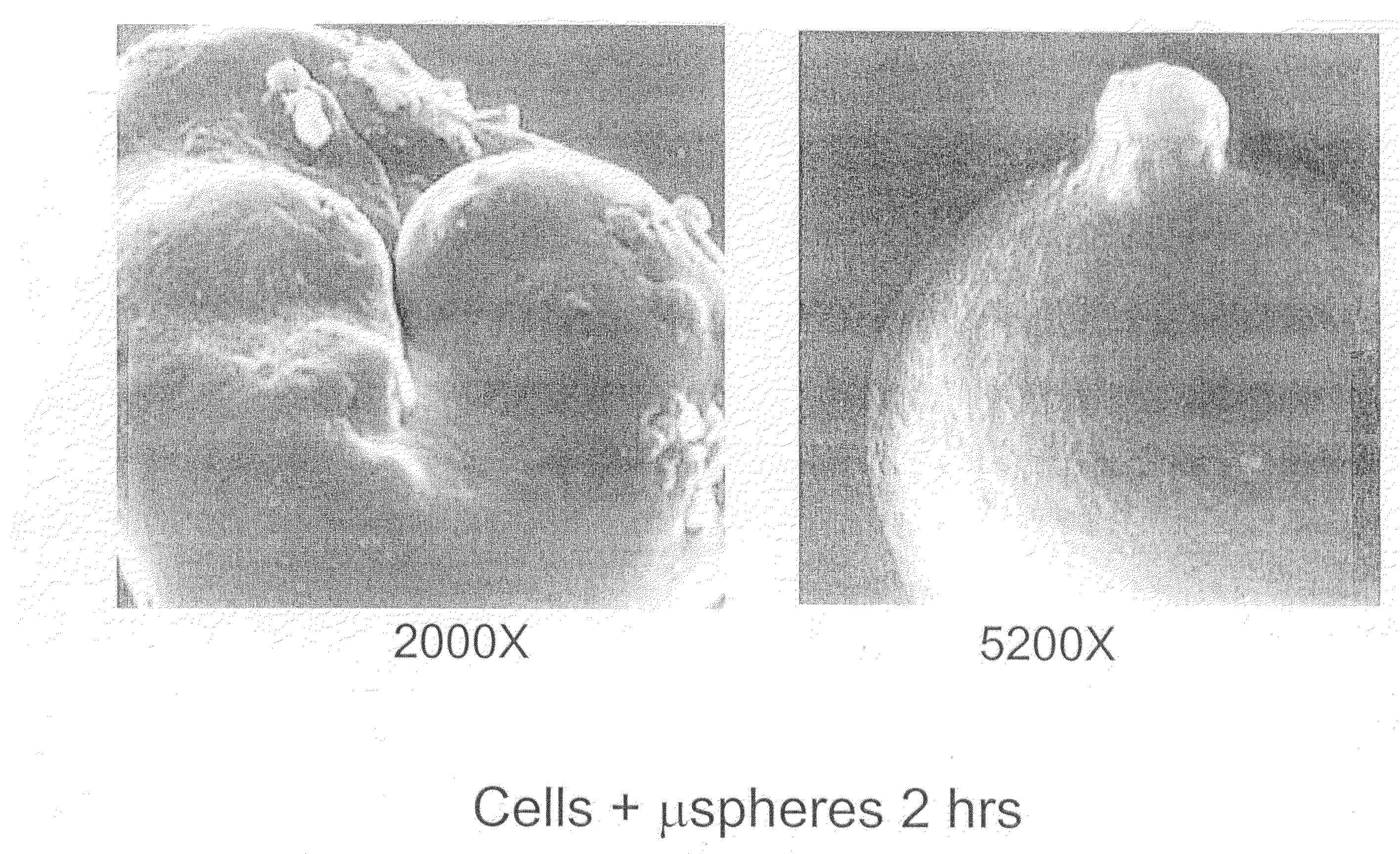
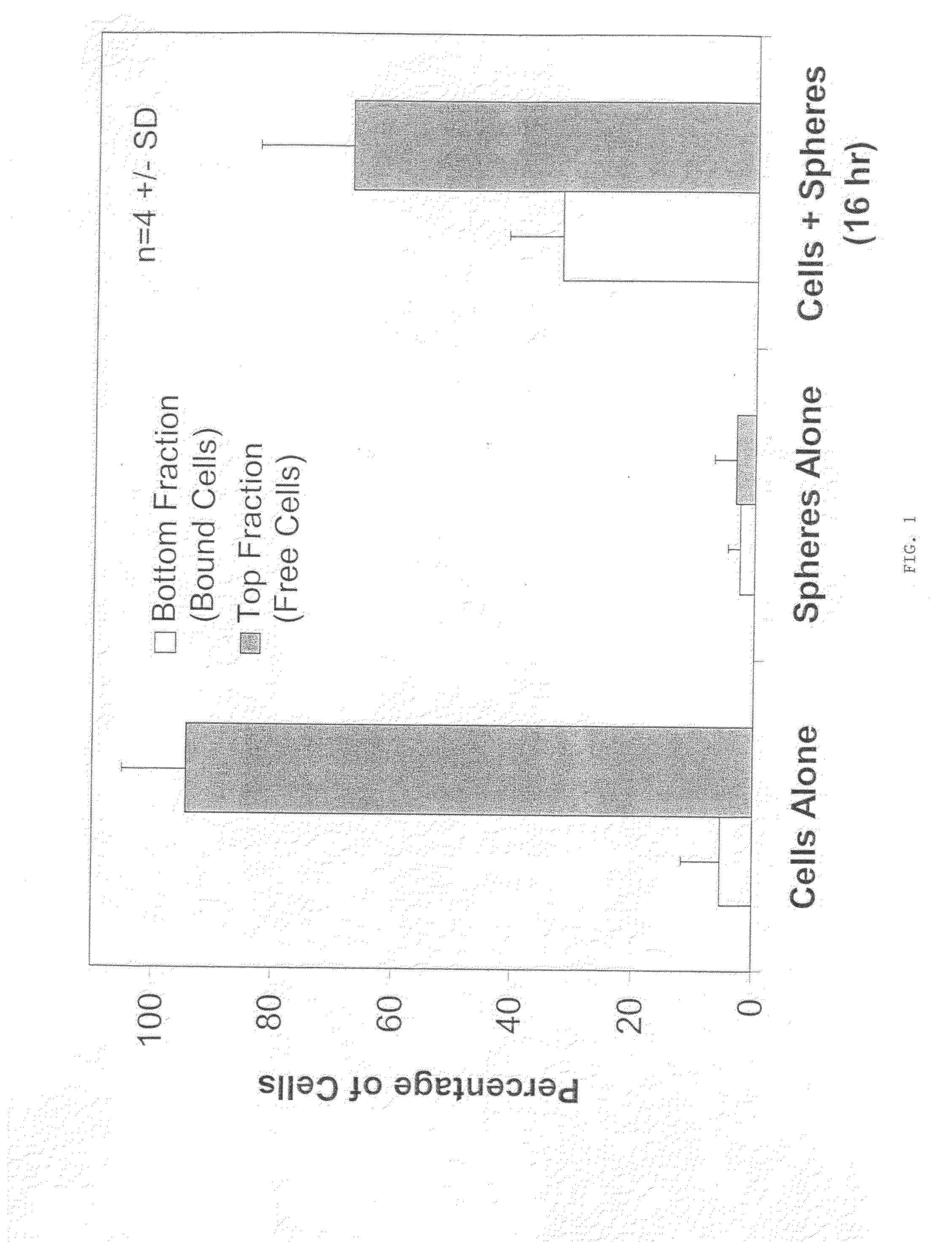
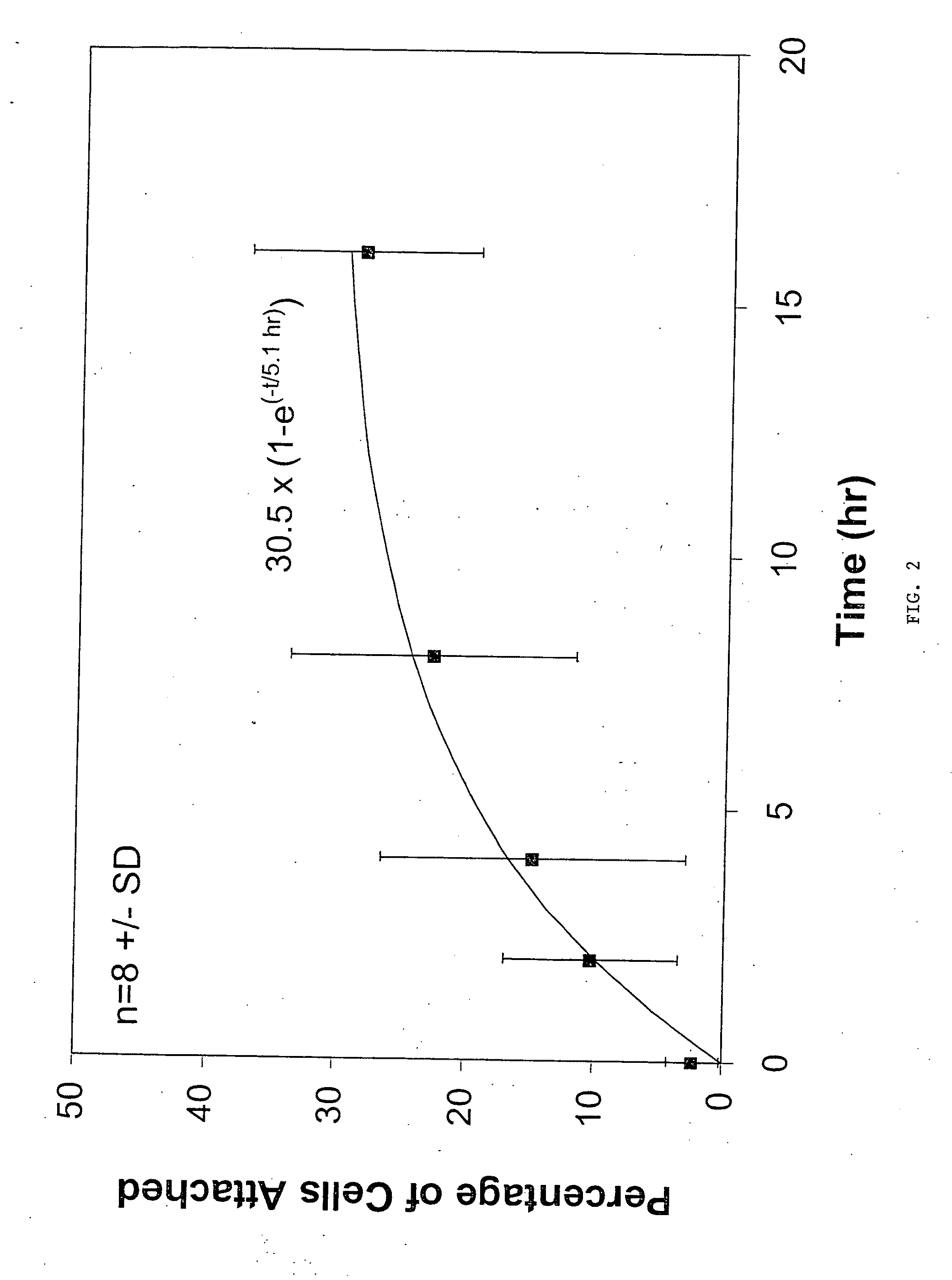
[0098]Chondrocytes were mixed with 3.57 mg / mL of PLG (50:50 L:G) microparticles, prepared as described above, to a final concentration of 1×10⁶ cells / mL. Control groups included cells alone and microparticles alone at the same concentrations as described above. The mixture of cells and microparticles contained phosphate buffered saline and cell culture medium.

[0099]The combined cell / microparticle suspension and controls were incubated on a shaker plate at 37° C. At 0, 2, 4, 16 hours, 1 mL samples were removed and assayed to determine the amount of cells which had adhered to the microparticles. In order to determine the amount of cells which had adhered to the microparticles, each sample was loaded onto a 3 mL histopaque density gradient and centrifuged for 5 min at 5000 rpm. The unattached cells were removed by decanting the less dense media fraction on top of the histopaque (approximately 1 mL). The remaining portion (approximately 3 mL) con...


PUM

PropertyMeasurementUnit
concentrationaaaaaaaaaa

Abstract

The invention relates to an improved method for administering live cells to a patient and compositions useful in the method. The composition comprises live cells and biocompatible, biodegradable polymer microparticles. The cells and microparticles of the cell / microparticle composition can be contacted immediately prior to administration, or can be contacted in culture for a specified period of time prior to administration. In the method of the invention, an effective amount of the cell / microparticle composition is administered to a patient in need thereof by injection to a treatment site of the patient to provide a therapeutic effect in the patient. The therapeutic effect can be, for example, the formation of new tissue at the treatment site, or the production and secretion of a biologically active secretory molecule at the treatment site. The therapeutic effect resulting from injection of the cell / microparticle composition into a treatment site, is determined by the type of cell present in the composition. The composition comprising lives cells and biocompatible, biodegradable polymer microparticles can further comprise a biologically active agent. In a preferred embodiment, the biologically active agent is incorporated into the microparticle. The biologically active agent can be, for example, factors which modulate cell growth.

Description

RELATED APPLICATION(S)[0001]This application is a continuation of U.S. application Ser. No. 10 / 784,908, filed Feb. 23, 2004, which is a continuation of U.S. application Ser. No. 09 / 612,744, filed Jul. 10, 2000, now U.S. Pat. No. 6,719,970, issued Apr. 13, 2004. The entire teachings of the above application(s) are incorporated herein by reference.BACKGROUND OF THE INVENTION[0002]Implantable polymeric materials capable of being degraded and absorbed by the body have been used in medicine for many years. For example, implantable devices can be pre-seeded with a desired cell type, and used as structural supports or scaffolds for guiding tissue regeneration. One example is the regeneration of cartilage tissue using a degradable fiber mesh pre-seeded with chondrocytes as described in U.S. Pat. No. 5,041,138 to Vacanti et al. However, implantation of the scaffolds requires surgical intervention which presents disadvantages, such as the risk of infection and the need for invasive and painfu...

Claims


Application Information

Patent Timeline
Patent Type & Authority Applications(United States)
IPC IPC(8): A61K35/12A61K9/14A61K35/32A61K35/39A61P3/10A61P19/04A61K35/30C12N5/077
CPCA61K35/32A61K35/30C12N2533/40C12N5/0655C12N2531/00A61K35/39A61P19/04A61P3/10
Inventor COSTANTINO, HENRY R.BONASSAR, LAWRENCE J.TRACY, MARK A.
Owner COSTANTINO HENRY R
Learn More