Broadband illuminator for oximetry, hemoglobin, met-hemoglobin, carboxy-hemoglobin, and other blood component monitoring

a broadband illuminator and oximetry technology, applied in the field of broadband illuminator for oximetry, hemoglobin, methemoglobin, carboxyhemoglobin, and other blood component monitoring, can solve the problems of difficult discrimination of the source of changes in tissue oxygenation, device not suitable for somatic monitoring, and device not allowing mutual or computational determination of difference-weighted value, etc., to achieve more sensitive detection of local conditions. , the effect o

Inactive Publication Date: 2009-02-19
J FITNESS LLC
View PDF57 Cites 10 Cited by
  • Summary
  • Abstract
  • Description
  • Claims
  • Application Information

AI Technical Summary

Benefits of technology

[0008]The inventors have discovered that certain diseases (vascular ischemia, cancer) are frequently localized, and by comparing at least two somatic values—either multiple sites or times—within the body, resulting in a more sensitive detection of such local conditions.
[0009]A salient feature of the present invention is that the detection and treatment of diseases such as somatic ischemia or cancer is aided by use of at least two measurements—either by multiple somatic sensors monitoring at least two nearby or distant regions or by dual measurements made by a single sensor over space or time—allowing a direct comparison of these different spectroscopic values by mutual inspection or computation.

Problems solved by technology

However, discriminating the source of changes in tissue oxygenation can be difficult, considering values at each site individually.
The great majority of such known devices and methods monitor only at one site (U.S. Pat. No. 6,662,033, WO / 2003 / 003914); such devices do not allow for mutual or computational determination of a difference-weighted value.
Further, the '065 patent teaches that it is the unique, hemispheric structure of the brain that allows the device of '065 to operate, and thus the device would not be suitable for somatic monitoring.
However, again no computational comparison is taught, and one of these values is determined through invasive blood sample, not from spectrophotometric measurement of tissue itself.
All of the above devices are limited to being single measures of oxygenation, are limited or optimized by design or omission to non-somatic tissue, and / or do not allow direct and near-simultaneous mutual comparison or computational processing of at least two somatic values obtained by spectrophotometric measures.
None of the prior devices or methods allow for a difference-weighted spectroscopy that facilitates simultaneous or near-simultaneous comparison of spectroscopic values from two somatic regions or sites by inspection or computation.
Such a system has not been previously described, nor successfully commercialized.

Method used


Image

  • Broadband illuminator for oximetry, hemoglobin, met-hemoglobin, carboxy-hemoglobin, and other blood component monitoring
  • Broadband illuminator for oximetry, hemoglobin, met-hemoglobin, carboxy-hemoglobin, and other blood component monitoring
  • Broadband illuminator for oximetry, hemoglobin, met-hemoglobin, carboxy-hemoglobin, and other blood component monitoring

Examples


example 1

Simultaneous Two-Site Two-Organ Somatic Difference Monitoring

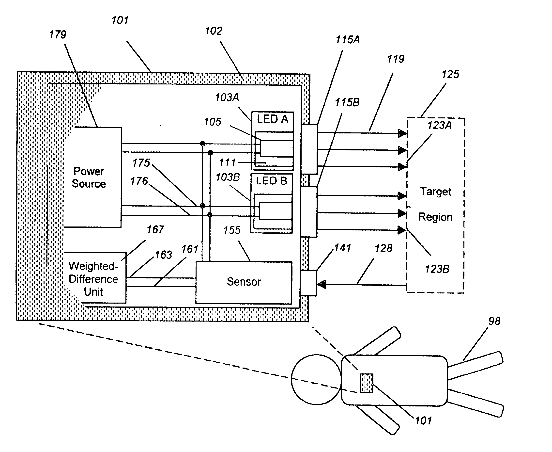
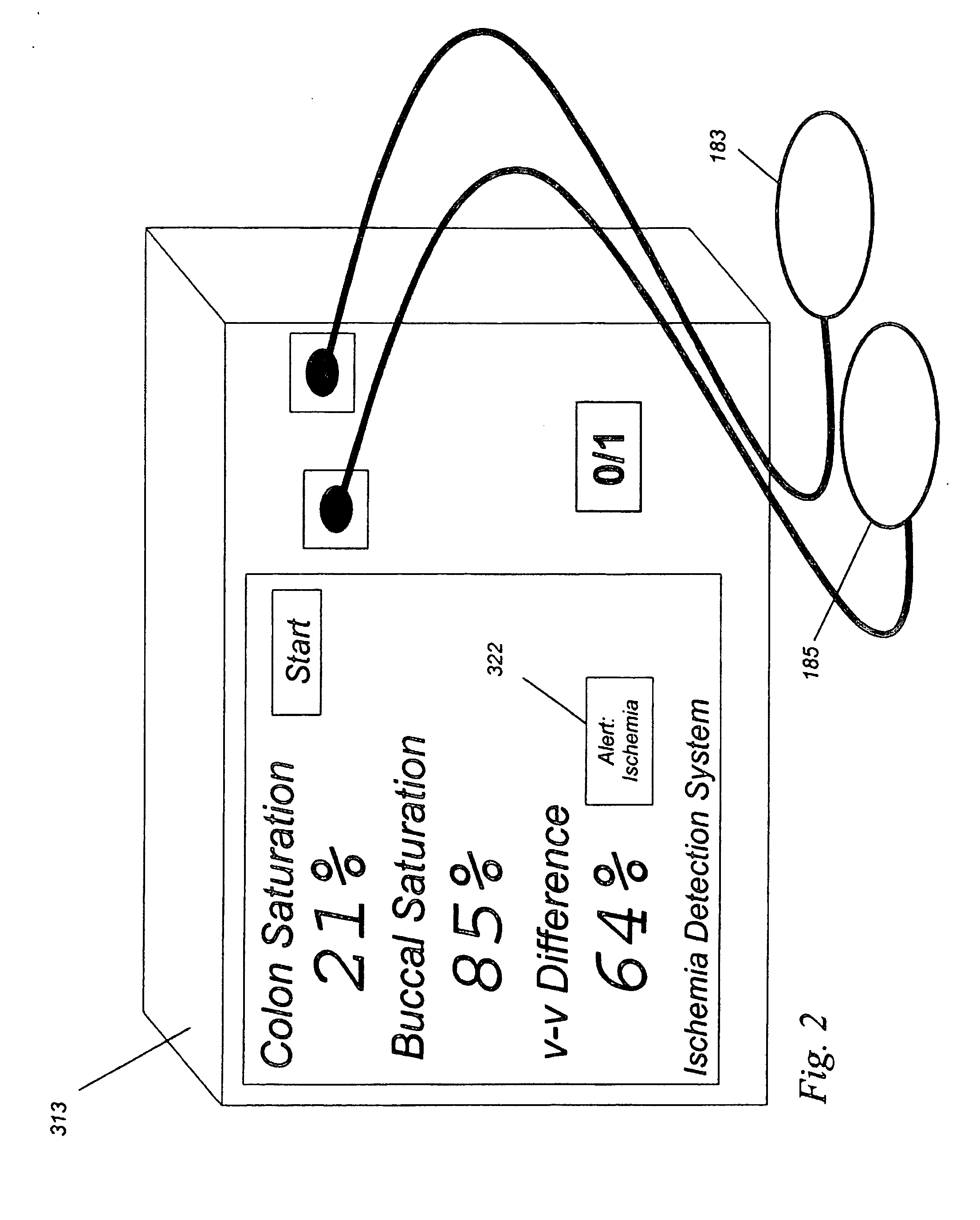
[0047]In this example, a clinical application related to ischemia is described. Here, a surgeon is repairing the aorta. There are several reasons why the local tissue oxygenation may fall. For example, the patient is under anesthesia, and a general depression (reduction) of cardiac output may occur. If so, the delivery of oxygen to all parts of the body will fall. On the other hand, if the blood vessel supplying the colon, which arises in part from the aorta, is occluded, then the saturation to the colon will fall, but not the saturation to the cheek. Therefore, by looking at the saturation of both the cheek and colon at substantially the same time, or by displaying a difference between the two values, the cause of the drop in local oxygenation may be determined to be either local and due to the vascular repair (e.g., large difference, in this case the absolute value of |Δ saturation|>10%) which is an indication of local i...

example 2

Simultaneous Two-Site Single-Organ Somatic Difference Monitoring

[0051]In the example above, two different organs were studied. In this example, the monitoring of a single organ, the breast, is described. It is toward this Example that the embodiment of FIG. 1 is directed.

[0052]In breast cancer, the detection of angiogenesis, the proliferation of new blood vessels, is a key feature of cancer that lets the cancer gain the ability to grow and spread. However, the background variation in blood content in the breast between women of different ages and breast composition makes the use of a single-site blood-content threshold less useful than it could otherwise be. That is, the range of normal blood content in breast tissue between different women is so large that the increase in blood due to cancer can be lost in that broad range.

[0053]To illustrate this, consider data from women with breast cancer. By looking at the difference measurement of the oxygenation at one location on the breast ...

example 3

Multi-Site Single-Organ Somatic Difference Monitoring

[0055]In the above example, pairs of data were taken, one pair at a time. In this example, instead of plotting values from a single pair, embodiments of the present invention provide for plotting real time difference values from many measures at many sites.

[0056]Again, using data from human subjects with and without breast cancer, the following table can be generated. Such differences can be found by having a difference in spatial separation at two points, as shown as the difference (delta) values at 5 sites labeled A-E on each subject, as follows:

TABLE 3The Spatial difference at multiple sites by plotting differences,reduces the noise in breast tissue saturation, and allows simpledetection of tumor near site C of Patient Tumor 4, in which thesaturation difference has a negative then positive deflection(or vice-versa) during scanning.PatientΔ Site AΔ Site BΔ Site CΔ Site DΔ Site ENormal 12%4%−3%5%−3%Normal 20%−2%3%2%−3%Normal 31%4...


PUM


Abstract

An ischemia detection system in two or more somatic measures, collected simultaneously or near-simultaneously, are provided for direct or computational comparison, in which light from light source A (103A) and source B (103B) is detected by a sensor (155), and a difference-weighted value is determined (167), thereby enhancing the value of the spectroscopic measurements over values taken individually and singly.

Description

CROSS REFERENCE TO RELATED APPLICATIONS[0001]This application is a continuation-in-part of U.S. patent application Ser. No. 11 / 451,681 filed on Jun. 12, 2006, relating to the detection of local tissue ischemia, which is a continuation-in-part of U.S. patent application Ser. No. 10 / 651,541 filed on Aug. 29, 2003, now U.S. Pat. No. 7,062,306, which is a continuation of U.S. patent application Ser. No. 10 / 119,998 filed on Apr. 9, 2002, now U.S. Pat. No. 6,711,426, the disclosures of all of which are incorporated in full by reference.FIELD OF THE INVENTION[0002]The present invention relates to devices and methods for providing, simultaneously or near-simultaneously, spectroscopic analysis from more than one somatic site, and more particularly relates to the determination of a difference-weighted analysis wherein the near-simultaneous determination of two (or more) spectroscopically-determined somatic oxygenation saturation values is performed in a manner allowing for the direct and near...

Claims


Application Information

Patent Timeline
Patent Type & Authority Applications(United States)
IPC IPC(8): A61B5/1455
CPCA61B5/14551A61B5/0261
Inventor BENARON, DAVID A.PARACHIKOV, ILLIAN H.FIERRO, MICHAEL R.
Owner J FITNESS LLC
Why Eureka
Try Eureka