
Method and apparatus for imaging target components in a biological sample using permanent magnets

a technology of permanent magnets and target components, which is applied in the direction of filtration separation, instruments, separation processes, etc., can solve the problems of low cost, inconsistent reliability, and inability to provide rapid and reliable methods

Active Publication Date: 2009-03-05
MENARINI SILICON BIOSYSTEMS SPA
View PDF13 Cites 35 Cited by
  • Summary
  • Abstract
  • Description
  • Claims
  • Application Information

AI Technical Summary

Benefits of technology

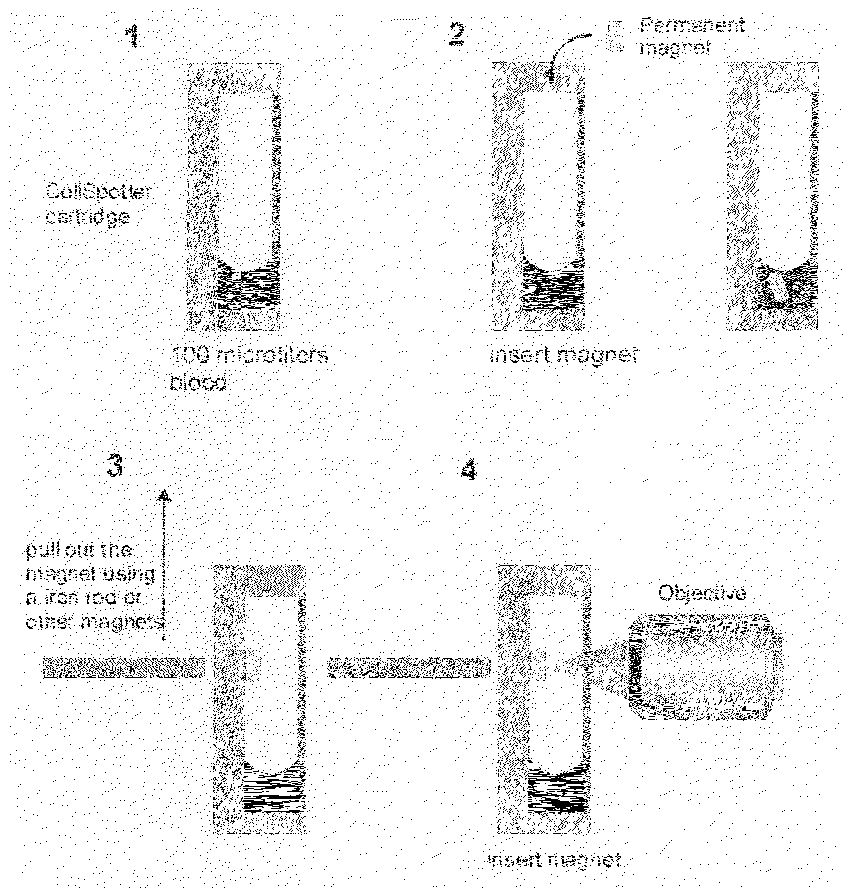
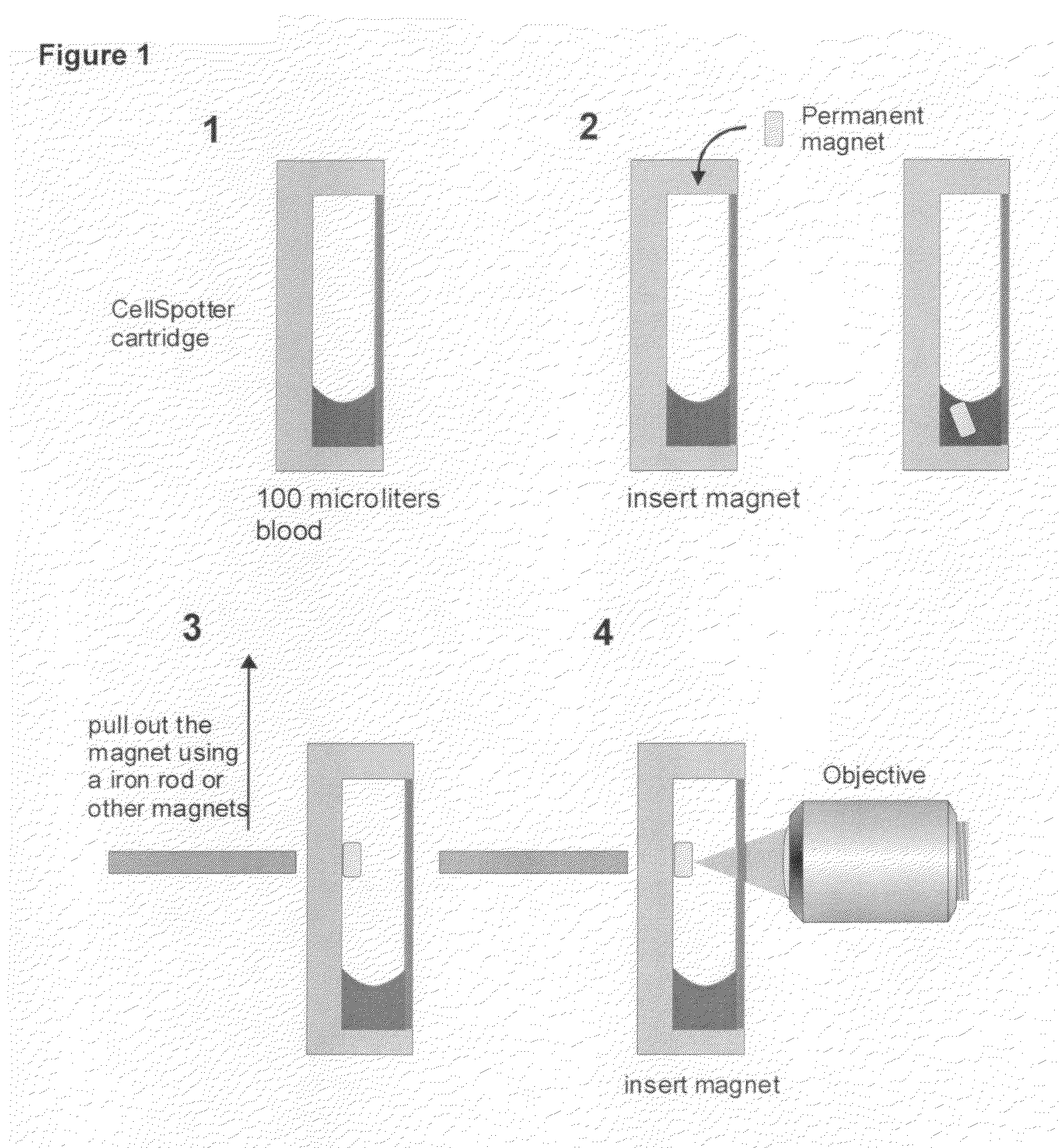
[0010]More specifically, the present invention provides the apparatus, methods, and kits for diagnosing disease disorders after immunomagnetic imaging. After obtaining a whole blood sample from a patient, a small permanent magnet is added to the whole blood sample. Unlike previously described CellSpotter Magnest configuration, a small NdFeB magnet is directly added to a sample container, for example the CellSpotter cartridge U.S. Pat. No. 6,______, with 100 ul of CD4 immunomagnetically labeled and fluorescently stained AO whole blood. After 10 minutes the small permanent magnet is pulled out of the sample using an iron rod or another magnet. The magnet is positioned within the container to allow for image analysis.

Problems solved by technology

Currently available methods do not provide a rapid, low cost and consistently reliable means for assessing a target population of cells by flow or image cytometry.

Method used


Image

  • Method and apparatus for imaging target components in a biological sample using permanent magnets
  • Method and apparatus for imaging target components in a biological sample using permanent magnets
  • Method and apparatus for imaging target components in a biological sample using permanent magnets

Examples


Embodiment Construction

[0015]Immunomagnetic isolation, enrichment, and analysis in blood combines immunomagnetic enrichment technology and immunofluorescent labeling technology with an appropriate analytical platform after initial blood draw. The associated test has the sensitivity and specificity to detect rare cells in a sample of whole blood with the utility to investigate their role in the clinical course of the disease such as malignant tumors of epithelial origin.

[0016]With this type of technology, circulating tumor cells (CTC) have been shown to exist in the blood in detectable amounts Image cytometric analysis such that the immunomagnetically enriched sample is analyzed by the Cell Spotter® System utilizes a fluorescence-based microscope image analysis system, which in contrast with flowcytometric analysis permits the visualization of events and the assessment of morphologic features to further identify objects (U.S. Pat. No. 6,______).

[0017]The CellSpotter® System refers to an automated fluoresce...


PUM

PropertyMeasurementUnit
heightaaaaaaaaaa
diameteraaaaaaaaaa
widthaaaaaaaaaa

Abstract

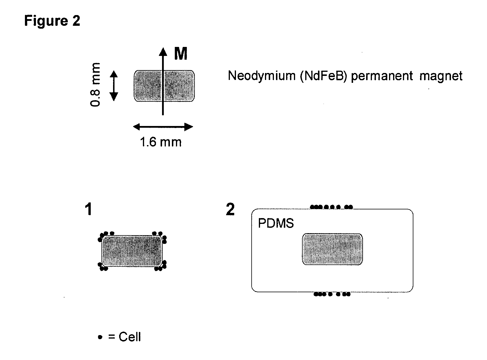
A system for enumeration of cells in fluids by image cytometry is described for assessment of target populations such as leukocyte subsets in different bodily fluids or bacterial contamination in environmental samples, food products and bodily fluids. Briefly, all cells in a biological sample are fluorescently labeled, but only the target cells are also magnetically labeled. A small, permanent magnet is inserted directly into the chamber containing the labeled sample. The magnets are coated with PDMS silicone rubber to provide a smooth and even surface which allows imaging on a single focal plane. The cells are illuminated and the images of the fluorescent light emitted by the target cells are captured by a CCD camera. Image analysis performed with a novel algorithm provides a count of the cells on the surface that can be related to the target cell concentration of the original sample.

Description

BACKGROUND [0001]1. Field of the Invention[0002]The invention relates generally to imaging target components in a biological sample. More specifically, methods and apparatus are described that provide for the positive selection of target cells in a blood sample. Small permanent magnets are added directly to a blood sample containing CD4 immuno-magnetic labeled fluorescently stained AO whole blood.[0003]2. Background Art[0004]The use of immunomagnetic separation technology provides greater sensitivity and specificity in the detection of target entities in blood for example, but not limited to, intact circulating cancer cells and endothelial cells. This simple and sensitive diagnostic tool, as described (U.S. Pat. No. 6,365,362; U.S. Pat. No. 6,551,843; U.S. Pat. No. 6,623,982; U.S. Pat. No. 6,620,627; U.S. Pat. No. 6,645,731; WO 02 / 077604; WO03 / 065042; and WO 03 / 019141) can be used in the present invention to correlate the statistical survivability of an individual patient based on a...

Claims


Application Information

Patent Timeline
Patent Type & Authority Applications(United States)
IPC IPC(8): C12Q1/06
CPCG01N33/54326G01N2333/70514G01N33/56972
Inventor TIBBE, ARIAN G.J.TERSTAPPEN, LEON W.M.M.
Owner MENARINI SILICON BIOSYSTEMS SPA
Learn More