Anatomical modeling from a 3-d image and a surface mapping

Active Publication Date: 2009-06-11
BIOSENSE WEBSTER INC
View PDF26 Cites 134 Cited by
  • Summary
  • Abstract
  • Description
  • Claims
  • Application Information

AI Technical Summary

Benefits of technology

[0061]There is further provided, in accordance with an embodiment of the present invention, a computer software product for medical imaging, the product including a computer-readable medium in which program instructions are stored, which instructions, when read by a computer, c

Problems solved by technology

Ultrasound imaging is a relatively convenient mode of real-time imaging, though the resolution of real-time ultrasound images is generally no

Method used


Image

  • Anatomical modeling from a 3-d image and a surface mapping
  • Anatomical modeling from a 3-d image and a surface mapping
  • Anatomical modeling from a 3-d image and a surface mapping

Examples


Embodiment Construction

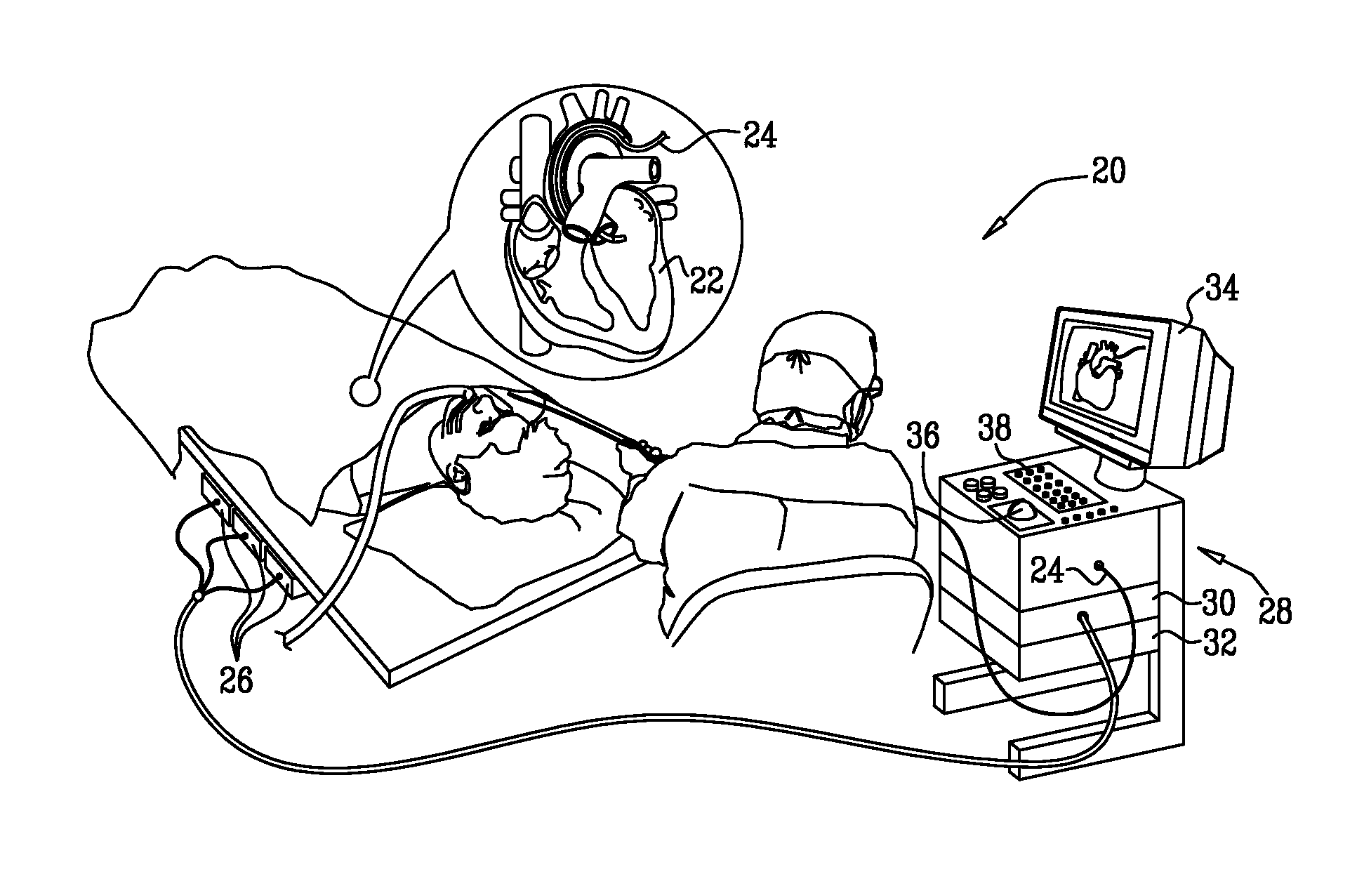
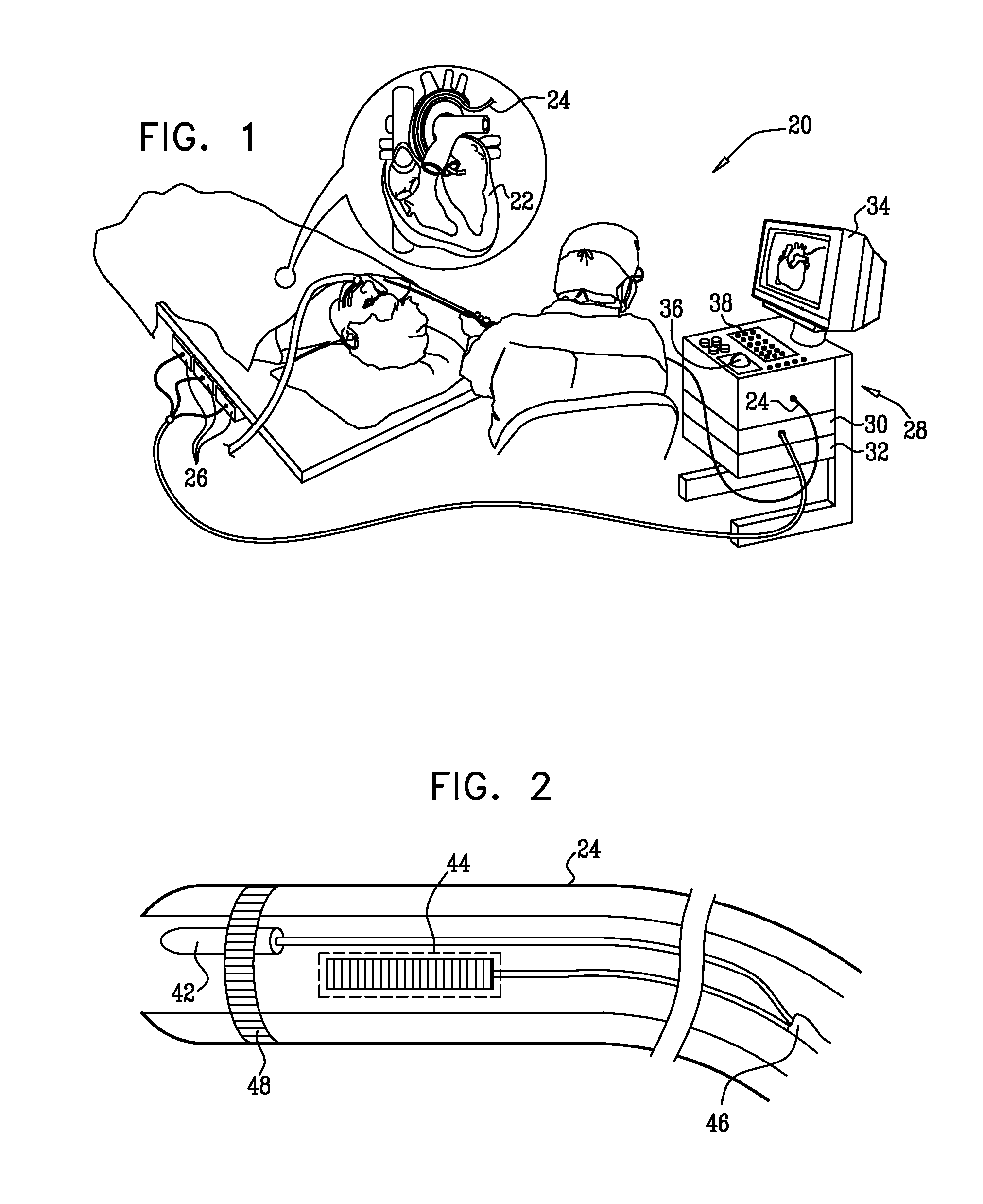
[0067]FIG. 1 is a schematic, pictorial illustration of a system 20 for imaging and mapping a target structure, such as a heart 22 of a patient, in accordance with an embodiment of the present invention. (Hereinbelow, the term “target structure” may refer to a chamber of the heart, in whole or in part, or other body cavity, or to a particular wall, surface, blood vessel or other anatomical feature. Although the embodiments described herein refer particularly to structures in and around the heart, the principles of the present invention may similarly be applied, mutatis mutandis, in imaging of bones, muscles and other organs and anatomical structures.)

[0068]The system comprises a catheter 24, which is inserted by a physician into a chamber of the heart. Typically, catheter 24 is a position-sensing, ultrasound probe, which is configured to perform functions that include anatomical mapping and ultrasound imaging. Mapping and ultrasound imaging capabilities of catheter 24 are further des...


PUM


Abstract

A method of medical imaging includes creating an anatomical map of an inner wall of a cavity in a body of a subject by inserting a probe into the body and collecting data using the probe. A three dimensional (3-D) contour is delineated in a 3-D image of the cavity based on the map.

Description

FIELD OF THE INVENTION[0001]The present invention relates generally to medical imaging systems, and particularly to methods and systems for constructing three-dimensional views of internal organs.BACKGROUND OF THE INVENTION[0002]Three-dimensional (3-D) images of internal organs are useful in many catheter-based diagnostic and therapeutic applications, and real-time imaging is widely used during surgical procedures. Ultrasound imaging is a relatively convenient mode of real-time imaging, though the resolution of real-time ultrasound images is generally not as good as the resolution obtained from other imaging modalities, such as computerized tomography (CT) and magnetic resonance imaging (MRI).[0003]U.S. Pat. No. 6,106,466 to Sheehan, et al., whose disclosure is incorporated herein by reference, describes a method for generating a 3-D model of a heart from 2-D ultrasound images. Anatomical locations are manually identified in the 2-D images. A 3-D mesh serves as an archetypal heart s...

Claims


Application Information

Patent Timeline
IPC IPC(8): A61B5/00
CPCA61B5/042A61B8/0883A61B5/1076A61B5/7285A61B6/503A61B6/504A61B6/5247A61B6/541A61B8/0891A61B8/12A61B8/4472A61B8/4488A61B8/483A61B8/5238A61B2019/5289G06T7/0083G06T19/00G06T2207/10072G06T2207/20136G06T2207/20156G06T2207/30048A61B5/06A61B2090/364G06T7/12G06T7/181A61B5/061A61B5/283
Inventor ALTMANN, ANDRES CLAUDIOGOVARI, ASSAF
Owner BIOSENSE WEBSTER INC
Why Eureka
Try Eureka