System and Methods for Generating Three-Dimensional Images From Two-Dimensional Bioluminescence Images and Visualizing Tumor Shapes and Locations

a bioluminescence and image technology, applied in the field of three-dimensional image generation, can solve the problems of high cost, unsuitable for repeated and rapid imaging in laboratory settings, and ineffective efforts to generate useful 3d images with high resolution,

Inactive Publication Date: 2009-06-11
RUTGERS THE STATE UNIV
View PDF4 Cites 8 Cited by
  • Summary
  • Abstract
  • Description
  • Claims
  • Application Information

AI Technical Summary

Benefits of technology

[0009]The present invention relates to a system and methods for reconstructing 3D images from 2D bioluminescent images and visualizing tumor shapes and locations. A plurality of 2D bioluminescent images of a subject are acquired using any suitable bioluminescent imaging system. The 2D images are taken during a complete revolution about an axis of the subject, such that the imaging system is rotated about the subject by a predetermined angle between each image. Optionally, the imaging system could be held in a stationary position, the subject could be rotated through a complete revolution, and a plurality of images could be taken. After imaging, the 2D images are registered according to the rotation axis to align each image and to compensate for differences between adjacent images. After registration, corresponding features are identified between consecutive sets of 2D images. For each corresponding feature identified in each set of 2D images, an orthographic projection technique (or model) is applied, such that rays are projected through each point in the feature. The intersection points of the rays are plotted in a 3D image space. All of the 2D images are processed in the same manner, such that the resulting 3D image of a tumor is generated. The subject itself, as well as a container holding the subject, can also be rendered in the 3D image using the orthographic projection technique. The tumor can be registered with a reference image of the subject or an image of the container so that the precise shape and location of the tumor can be visualized, together with detailed anatomical structure information extracted from other imaging modalities such as microCT.

Problems solved by technology

Other imaging modalities, such as positron emission tomography (PET) and magnetic resonance imaging (MRI), are unsuitable for repeated and rapid imaging in laboratory settings.
Such efforts, however, have been largely ineffective in generating useful 3D images with high resolution.
The use of multiple CCD cameras to measure bioluminescence signals has been suggested, but this approach is expensive and requires careful calibration of multiple cameras.
As such, although BLI is a useful imaging modality for generating 2D images, there currently is no practical technique for rapidly generating three-dimensional (3D) images from a series of 2D BLI images.
In addition to the aforementioned limitations, existing BLI techniques do not allow for the rapid and accurate visualization of the physical shape and location of a tumor in a subject, in three dimensions.
While there has been extensive research on multi-modal image registration in the image processing literature (based on matching geometric features or the optimization of intensity-based energy functions), no practical, 3D visualization approach for BLI images has been developed.
Moreover, images generated by existing BLI techniques cannot be easily registered with 3D images generated by other imaging modalities (such as computed tomography (CT) and microCT) so that the physical shape and location of a tumor can be quickly discerned.

Method used


Image

  • System and Methods for Generating Three-Dimensional Images From Two-Dimensional Bioluminescence Images and Visualizing Tumor Shapes and Locations
  • System and Methods for Generating Three-Dimensional Images From Two-Dimensional Bioluminescence Images and Visualizing Tumor Shapes and Locations
  • System and Methods for Generating Three-Dimensional Images From Two-Dimensional Bioluminescence Images and Visualizing Tumor Shapes and Locations

Examples


Embodiment Construction

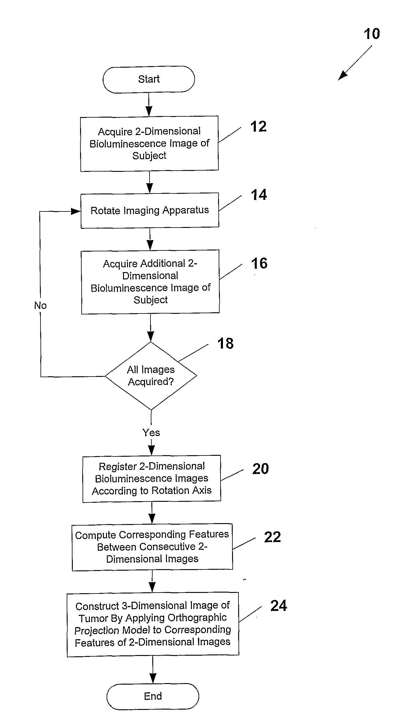
[0026]The present invention relates to a system and methods for generating (or reconstructing) 3D images from 2D bioluminescent images and visualizing tumor locations. A plurality of 2D bioluminescent images of a subject are acquired during a complete revolution of an imaging system about a subject, using any suitable bioluminescent imaging system. After imaging, the 2D images are registered according to the rotation axis to align each image and to compensate for differences between adjacent images. After registration, corresponding features are identified between consecutive sets of 2D images. For each corresponding feature identified in each set of 2D images, an orthographic projection technique (or model) is applied, such that rays are projected through each point in the feature. The intersection points of the rays are plotted in a 3D image space. All of the 2D images are processed in the same manner, such that a resulting 3D image of a tumor is generated. The 3D image can be reg...


PUM


Abstract

A system and methods for generating 3D images (24) from 2D bioluminescent images (22) and visualizing tumor locations are provided. A plurality of 2D bioluminescent images of a subject are acquired during a complete revolution of an imaging system about a subject, using any suitable bioluminescent imaging system. After imaging, the 2D images are registered (20) according to the rotation axis to align each image and to compensate for differences between adjacent images. After registration (20), corresponding features are identified between consecutive sets of 2D image (22). For each corresponding feature identified in each set of 2D images an orthographic projection model (24) is applied, such that rays are projected through each point in the feature. The intersection point of the rays are plotted in a 3D image of a tumor is generated. The 3D image can be registered with a reference image of the subject, so that the shape and location of the tumor can be precisely visualized with respect to the subject.

Description

RELATED APPLICATIONS[0001]This application claims the benefit of U.S. Provisional Application Ser. No. 60 / 715,610 filed Sep. 12, 2005, the entire disclosure of which is expressly incorporated herein by reference.BACKGROUND OF THE INVENTION[0002]1. Field of the Invention[0003]The present invention relates to the generation of three-dimensional images, and more particularly, to a system and methods for generating three-dimensional images from two-dimensional bioluminescence images and visualizing tumor shapes and locations.[0004]2. Related Art[0005]Bioluminescence imaging (BLI) is an emerging technique for sensitive and non-invasive imaging, which can be used for monitoring molecular events in living animals. Important applications of this imaging technique include gene therapy and cell trafficking studies. Unlike fluorescence-based optical imaging approaches which require an external source of light for excitation of fluorophores, BLI generates a two-dimensional (2D) view of gene exp...

Claims


Application Information

Patent Timeline
Patent Type & Authority Applications(United States)
IPC IPC(8): G06K9/00
CPCG06T7/0075G06T7/0012G06T7/593
Inventor METAXAS, DIMITRISBANERJEE, DEBABRATAHUANG, XIAOLEI
Owner RUTGERS THE STATE UNIV
Why Eureka
Try Eureka