Vascularized cardiac tissue and methods of producing and using same
a technology of cardiac tissue and vascularization, applied in the field of vascularized cardiac tissue, can solve the problems of poor prognosis for heart failure patients, affecting the clinical translation of these approaches, and progressing loss of ventricular function and heart failure developmen
- Summary
- Abstract
- Description
- Claims
- Application Information
AI Technical Summary
Benefits of technology
Problems solved by technology
Method used
Image
Examples
example 1
Generation of the Engineered Human Cardiac Tissue
[0180]To explore the ability of establishing a three-dimensional supportive environment for generation of a vascularized cardiac tissue, PLLA (50%) / PLGA (50%) biodegradable scaffolds were used. The PLGA was selected to allow relatively fast degradation (˜3 weeks) to facilitate cellular ingrowths, whereas the PLLA was chosen to provide mechanical support for the three-dimensional structure. Three cell culture combinations were evaluated: (1) scaffolds seeded with human embryonic stem cells differentiated into cardiomyocytes (hESC-CM; 4*105 cells) alone, (2) co-cultures comprised of hESC-CM (4*105 cells) and HUVEC or hESC-derived EC (hESC-EC9) (4*105 cells), and (3) a triple-cell culture comprised of hESC-CM, HUVEC or hESC-EC, supplemented with embryonic fibroblasts (EmF) (2*105-4*105 cells). The cells were seeded into the scaffolds together with matrigel to facilitate cell seeding and to keep the cells on the scaffolds.
[0181]The scaffo...
example 2
Vascularization of the Engineered Human Cardiac Tissue
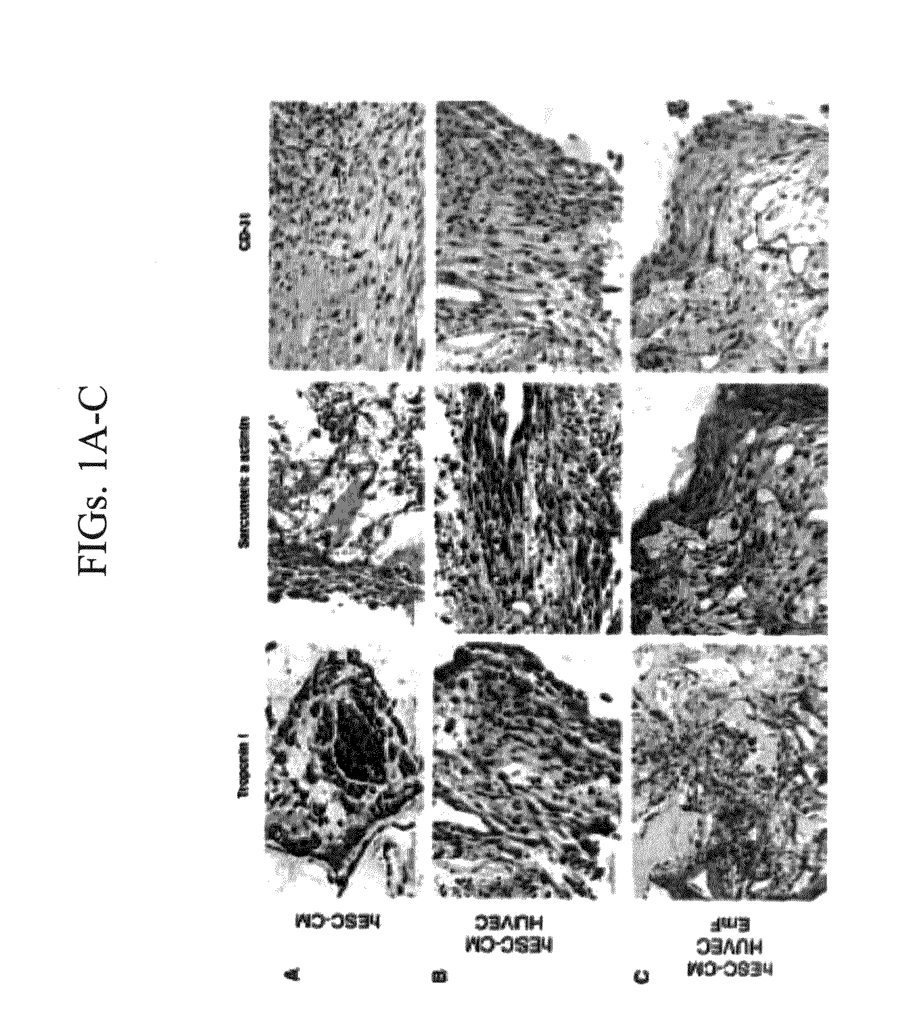
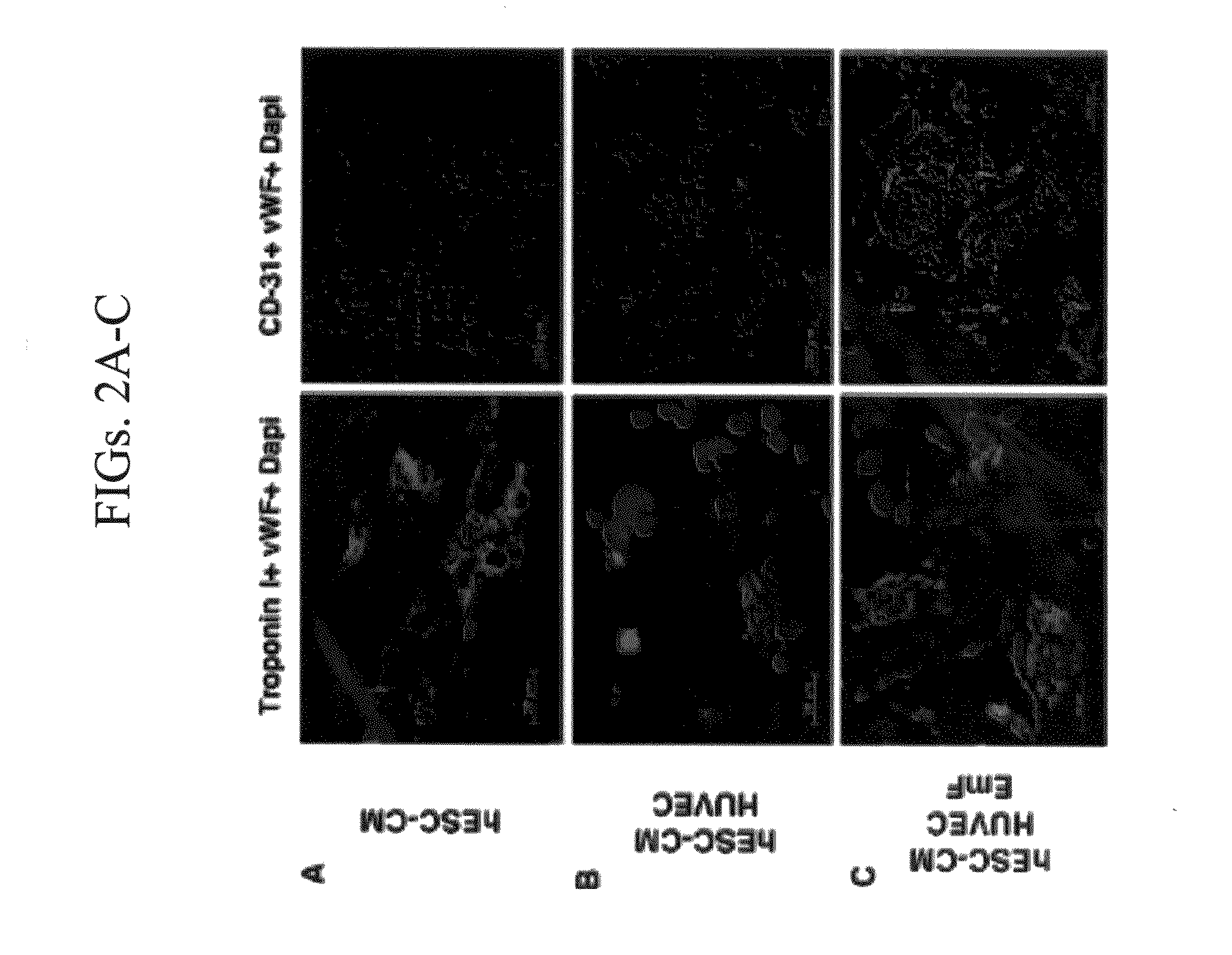
[0182]Scaffolds consisting of just hESC-CM contained only few vWF+ or CD-31+ cells (FIGS. 1A and 2A). The addition of HUVEC resulted in a significant increase in the quantity of endothelial cells (ECs) when compared to the scaffolds containing only hESC-CM (FIGS. 1B and 2B). Despite the increase in EC density, the EC did not organize into blood vessels and were mainly present as compact cell clusters (FIGS. 1B, 2B, and 3A).
[0183]The effects of adding embryonic fibroblasts (EmF) to the constructs containing hESC-CM and EC were examined. Examination of these tri-culture three-dimensional scaffolds revealed that the addition of EmF resulted in the generation of highly-vascularized engineered cardiac muscle (FIGS. 1C and 2C). Both immunohistochemical and immunofluorescence stainings demonstrated the organization of the EC into a condense network of vessels that was present within and in some cases also closely adjacent to the cardiac...
example 3
Expression of Angiogenic and Vasculogenic Factors
[0185]To evaluate the expression of key angiogenic and vasculogenic factors in the three-dimensional vascularized cardiac tissue, the expression of vascular endothelial growth factor (VEGF-A), platelet derived growth factor (PDGF-B), Angiopoietin 1 (Ang1), and basic fibroblast growth factor (bFGF) was assessed at the mRNA level. Similar to the histological quantification of the vascularization process, the RT-PCR analysis revealed increased gene expression of the angiogenic factors VEGF-A, PDGF-B and Ang1 in the tri-culture cardiac tissue (upon addition of EmF) (FIG. 4D). An increase in the bFGF mRNA levels in the tri-culture was not noted.
[0186]A major factor known to contribute to EC organization is the presence of pericytes or smooth muscle cells. It was therefore assessed whether the EmF in the tri-cultures differentiated into smooth muscle cells. Immunostainings for α-smooth muscle actin (SMA) demonstrated the presence of SMA+ ce...
PUM
 Abstract
Description
Claims
Application Information
 - R&D
- Intellectual Property
- Life Sciences
- Materials
- Tech Scout
- Unparalleled Data Quality
- Higher Quality Content
- 60% Fewer Hallucinations
Browse by: Latest US Patents, China's latest patents, Technical Efficacy Thesaurus, Application Domain, Technology Topic, Popular Technical Reports.
© 2025 PatSnap. All rights reserved.Legal|Privacy policy|Modern Slavery Act Transparency Statement|Sitemap|About US| Contact US: help@patsnap.com


