Method and apparatus for real-time hemodynamic monitoring

a hemodynamic monitoring and real-time technology, applied in the field of medical imaging with catheter-based sensors, can solve problems such as inaccurate data

Inactive Publication Date: 2009-07-02
ST JUDE MEDICAL ATRIAL FIBRILLATION DIV
View PDF3 Cites 64 Cited by
  • Summary
  • Abstract
  • Description
  • Claims
  • Application Information

AI Technical Summary

Benefits of technology

[0018]The control circuitry can generate pressure-volume loop data signals from the surface image data signals and intracardiac pressure data signals in real time, and can provide the pressure-volume lo...

Problems solved by technology

However, these catheters typically generate volume ...

Method used


Image

  • Method and apparatus for real-time hemodynamic monitoring
  • Method and apparatus for real-time hemodynamic monitoring
  • Method and apparatus for real-time hemodynamic monitoring

Examples


Embodiment Construction

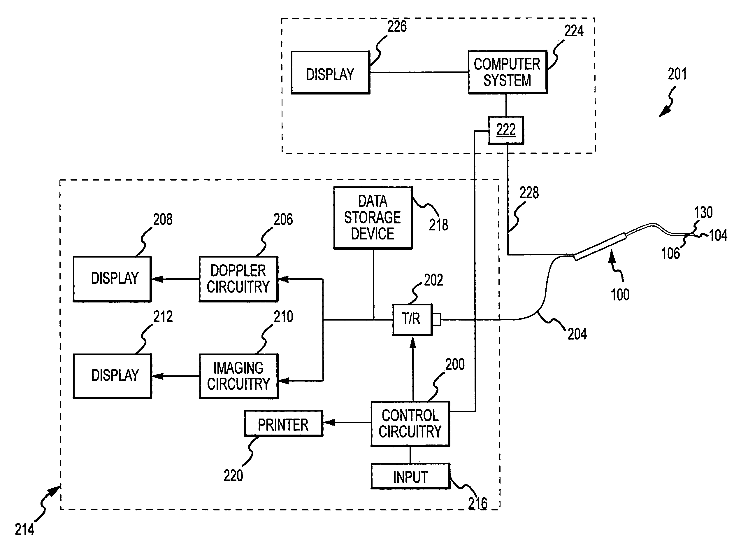
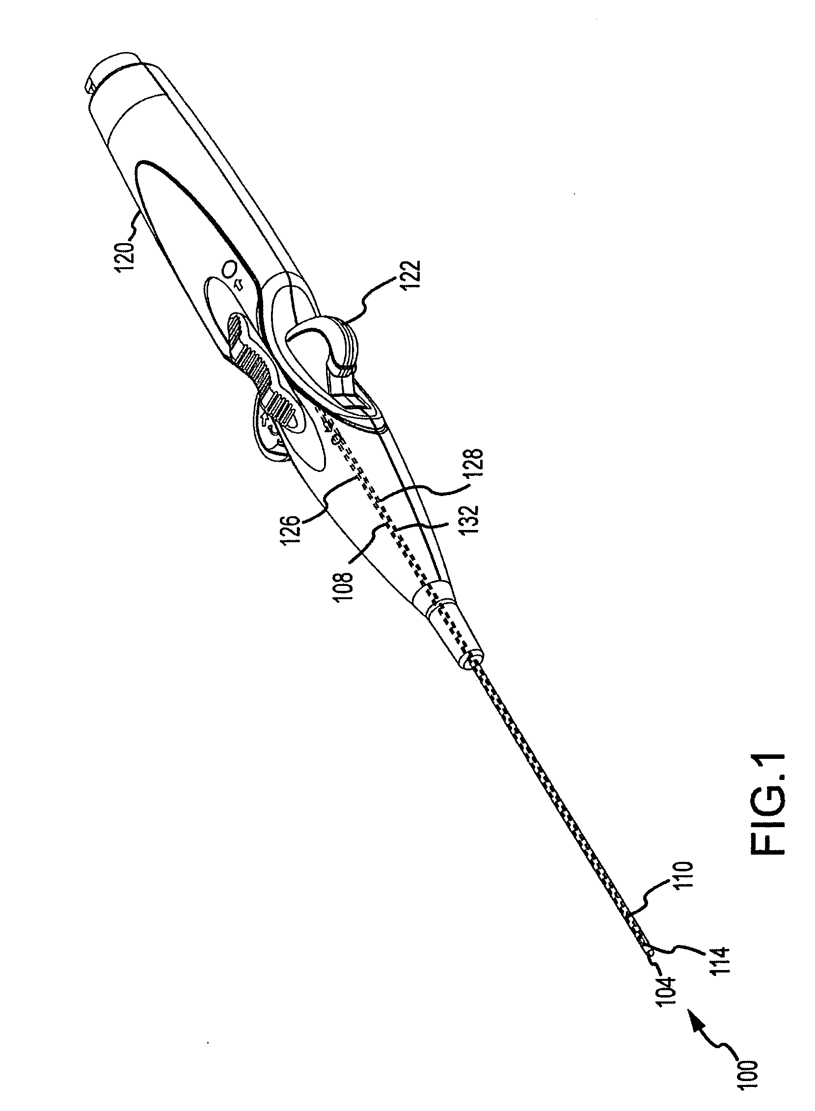
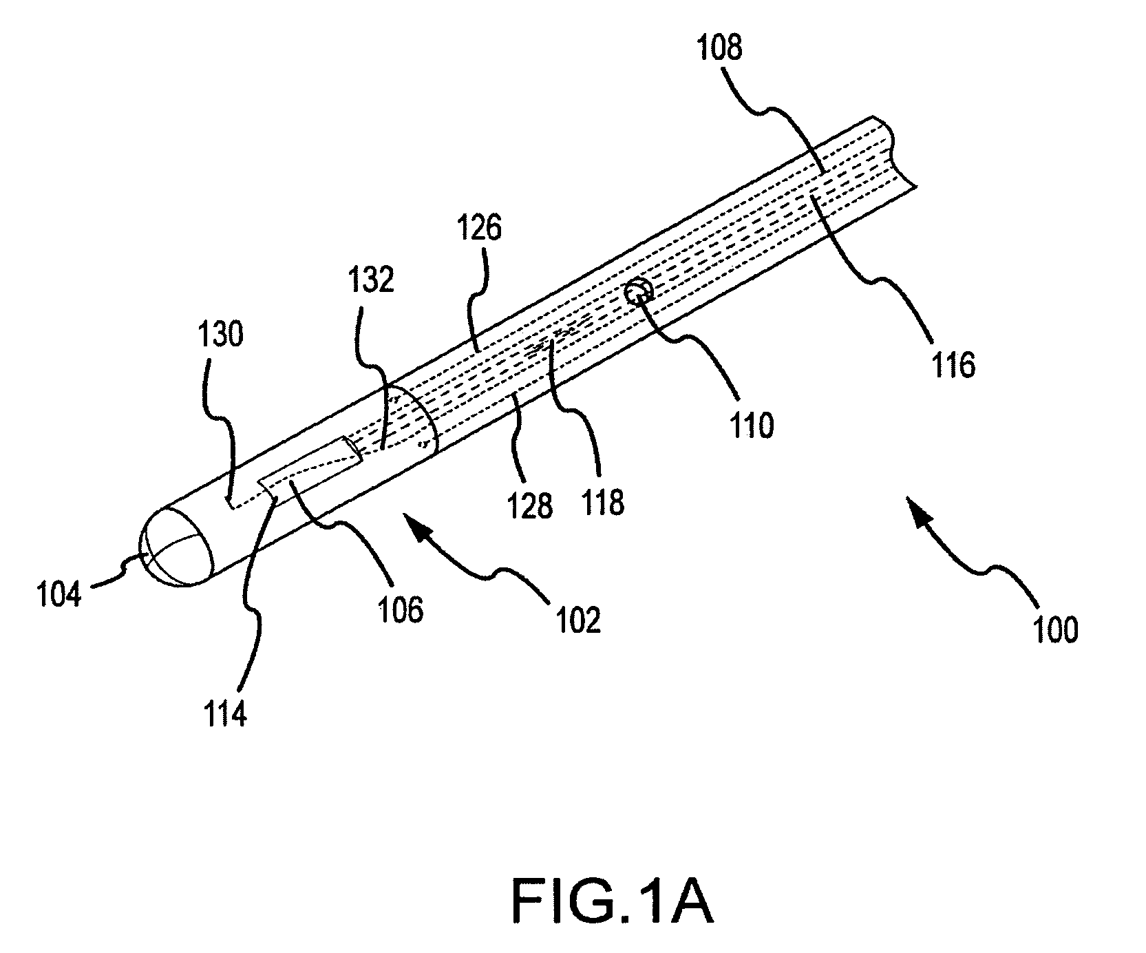
[0037]Referring to FIGS. 1 and 1A, catheter 100 includes an elongated flexible tubular catheter body 102 having a distal end 104. Catheter 100 includes near its distal end 104 an ultrasonic transducer 106 which is used to transmit ultrasound and receive resultant echoes to provide a field of view within which flowrates can be measured and features imaged. Transducer 106 can be any known transducer, including but not limited to single element, multiple element, cylindrical element, rotating element, linear array, curved array, circular array, vector phased array, linear phased array, circular phased array or any other mechanical array or dynamic array type that provides a flat two-dimensional tomogram or a volumetric three-dimensional image for intrabody operation within the field of ultrasound. Some examples of such transducers are outlined in N. Bom et al., Early and Recent Intraluminal Ultrasound Devices, ” International Journal of Cardiac Imaging, (1989) Vol. 4, pp. 79-88, incorp...


PUM


Abstract

The invention relates to an apparatus for monitoring hemodynamic performance of a cardiac chamber. In one embodiment, the apparatus takes real time measurements of the volume and pressure of the cardiac chamber and prepares a PV loop. The apparatus may include an intracardiac echocardiogram catheter with a pressure sensor positioned to measure intracardiac pressure when the distal end of the catheter is deployed in a cardiac chamber. The apparatus may further include control circuitry that receives heart wall surface image data signals from the ultrasound transducer and intracardiac pressure data signals from the pressure senor and generates pressure-volume loop data signals from the surface image data signals and intracardiac pressure data signals in real time.

Description

BACKGROUND OF THE INVENTION[0001]a. Field of the Invention[0002]The instant invention relates to medical imaging with catheter based sensors. In particular, the instant invention relates to a catheter system for measurement of cardiac chamber volume or collection of intra-cardiac pressure waveform data to implement a pressure volume loop system.[0003]b. Background Art[0004]Pressure-volume (PV) loops have become popular tools to characterize aspects of the performance of the heart. PV loops are derived from pressure and volume information found in the cardiac cycle. To generate a PV loop for the left ventricle, for example, the left ventricular pressure (LVP) is plotted against left ventricular (LV) volume at multiple time points during a complete cardiac cycle. When this is done, a PV loop is generated. Catheters designed to provide PV loops are known in the art, including those designed and manufactured by Millar Instruments. However, these catheters typically generate volume data ...

Claims


Application Information

Patent Timeline
IPC IPC(8): A61B5/02A61B8/08
CPCA61B5/0215A61B6/503A61B8/06A61B8/0883G06T2207/30048G06T7/0012G06T2207/10132G06T2207/20161A61B8/12
Inventor OLSON, ERIC S.
Owner ST JUDE MEDICAL ATRIAL FIBRILLATION DIV
Why Eureka
Try Eureka