Method and kit for detecting the early onset of renal tubular cell injury
a kidney and tubular cell technology, applied in the field of kidney tubular cell injury detection methods and kits, can solve the problems of delayed initiation, high mortality and morbidity, and high mortality
- Summary
- Abstract
- Description
- Claims
- Application Information
AI Technical Summary
Benefits of technology
Problems solved by technology
Method used
Image
Examples
example 1
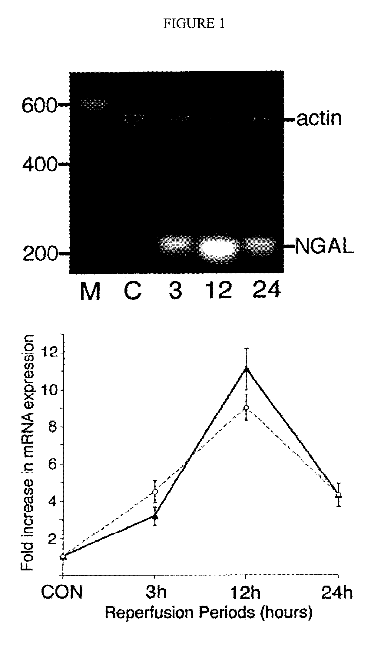
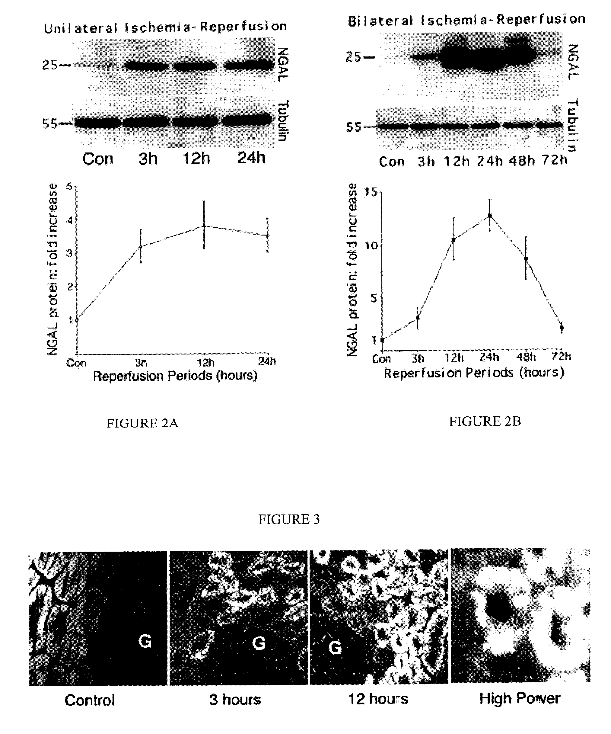
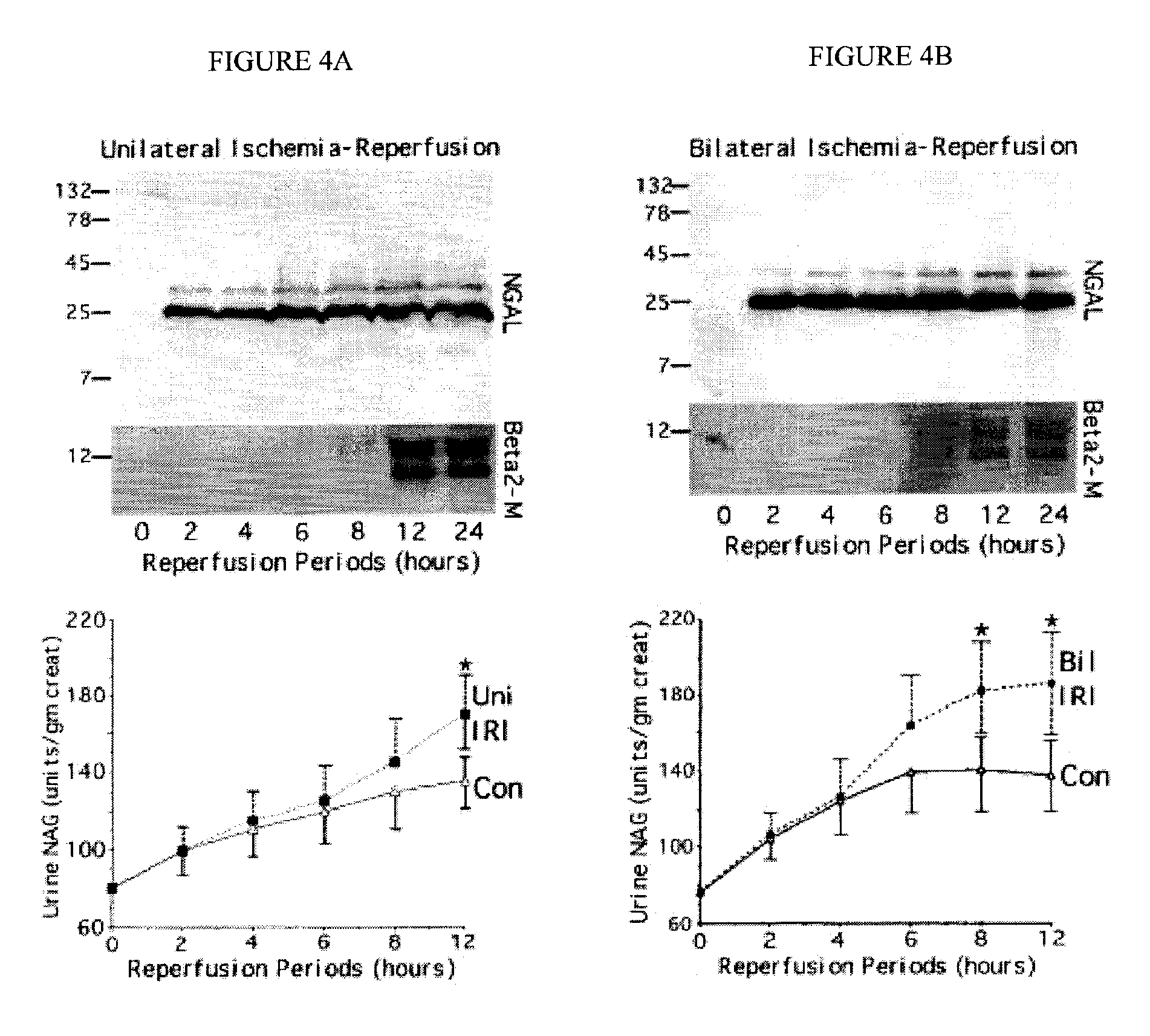
[0087]NGAL is a small protease-resistant, secreted polypeptide that is detectable in the urine. The marked upregulation of NGAL mRNA and protein levels has been shown in the early post-ischemic mouse kidney. NGAL protein expression was detected predominantly in proximal tubule cells, in a punctate cytoplasmic distribution reminiscent of a secreted protein. Indeed, NGAL was easily and rapidly detected in the urine (in the very first urine output) following ischemic injury in both mouse and rat models of ARF, at which time no leukocytic infiltration of the kidney was observed. The origin of NGAL from tubule cells was further confirmed in cultured human proximal tubule cells subjected to in vitro ischemic injury, where NGAL mRNA was markedly and promptly induced in the cells, and NGAL protein readily detectable in the culture medium within one hour of mild ATP depletion. Our results indicate that NGAL may represent a novel early urinary biomarker for ischemic renal injury.
Identificatio...
example 2
NGAL Protein is Easily Detected in the Urine Immediately after Induction of ARF in Rats
[0095]Since a debate exists regarding species differences in the responses to renal artery occlusion (10), we next examined the behavior of NGAL in a different animal model, namely a well-established rat model of renal ischemia-reperfusion injury. Using urinary creatinine concentrations to equalize for sample loading, NGAL was absent from the urine prior to rat renal ischemia. In marked contrast, NGAL was manifest as a 25 kDa immunoreactive peptide within 3 h of the injury (in the very first urine output following ischemia), as shown in FIG. 6. In comparison, the serum creatinine in this model of ischemic injury was elevated only after 24 h of reperfusion (not shown). Once again, NGAL was detectable in 1 μl of unprocessed urine and persisted for the entire duration examined (24 h of reperfusion).
example 3
NGAL mRNA is Induced in Cultured Human Proximal Tubule Cells after Early Mild Ischemia
[0096]In order to confirm the origin of NGAL from ischemic proximal tubule cells, we modified previously described protocols of in vitro ischemia by ATP depletion in cultured human proximal tubule cells (RPTEC). Incubation in 1 μm antimycin resulted in a mild partial ATP depletion to about 83±3% of control within 1 h, with a more gradual decrease to about 75±3% of control by 6 h (mean+ / −SD from four experiments). No morphological consequences of this mild ATP depletion were discernible. NGAL mRNA was just detectable in resting cells. However, following partial ATP depletion, a rapid and duration-dependent induction of NGAL mRNA was evident by RT PCR, as shown in FIG. 7.
NGAL Protein is Easily Detected in the Medium after Early Ischemia In Vitro:
[0097]We next examined NGAL protein expression in RPTEC cells and the culture medium following mild ATP depletion. NGAL protein was detectable in control RPT...
PUM
 Abstract
Description
Claims
Application Information
 - R&D
- Intellectual Property
- Life Sciences
- Materials
- Tech Scout
- Unparalleled Data Quality
- Higher Quality Content
- 60% Fewer Hallucinations
Browse by: Latest US Patents, China's latest patents, Technical Efficacy Thesaurus, Application Domain, Technology Topic, Popular Technical Reports.
© 2025 PatSnap. All rights reserved.Legal|Privacy policy|Modern Slavery Act Transparency Statement|Sitemap|About US| Contact US: help@patsnap.com


