Sensitometric response mapping for radiological images

a sensitometric and image technology, applied in the field of radiological images, can solve the problem of still further complexity of this problem

Inactive Publication Date: 2009-07-30
CARESTREAM HEALTH INC
View PDF4 Cites 17 Cited by
  • Summary
  • Abstract
  • Description
  • Claims
  • Application Information

AI Technical Summary

Benefits of technology

[0009]An advantage of the present invention is that it provides a signal mapping method that can be adapted for mapping image data between different imaging modalities.
[0010]These and other objects, features, and advantages of the pres...

Problems solved by technology

Because the types of signal values obtained from the receiver for these modalities are not the same, there is still further complexity to this problem than FIGS...

Method used


Image

  • Sensitometric response mapping for radiological images
  • Sensitometric response mapping for radiological images
  • Sensitometric response mapping for radiological images

Examples


Embodiment Construction

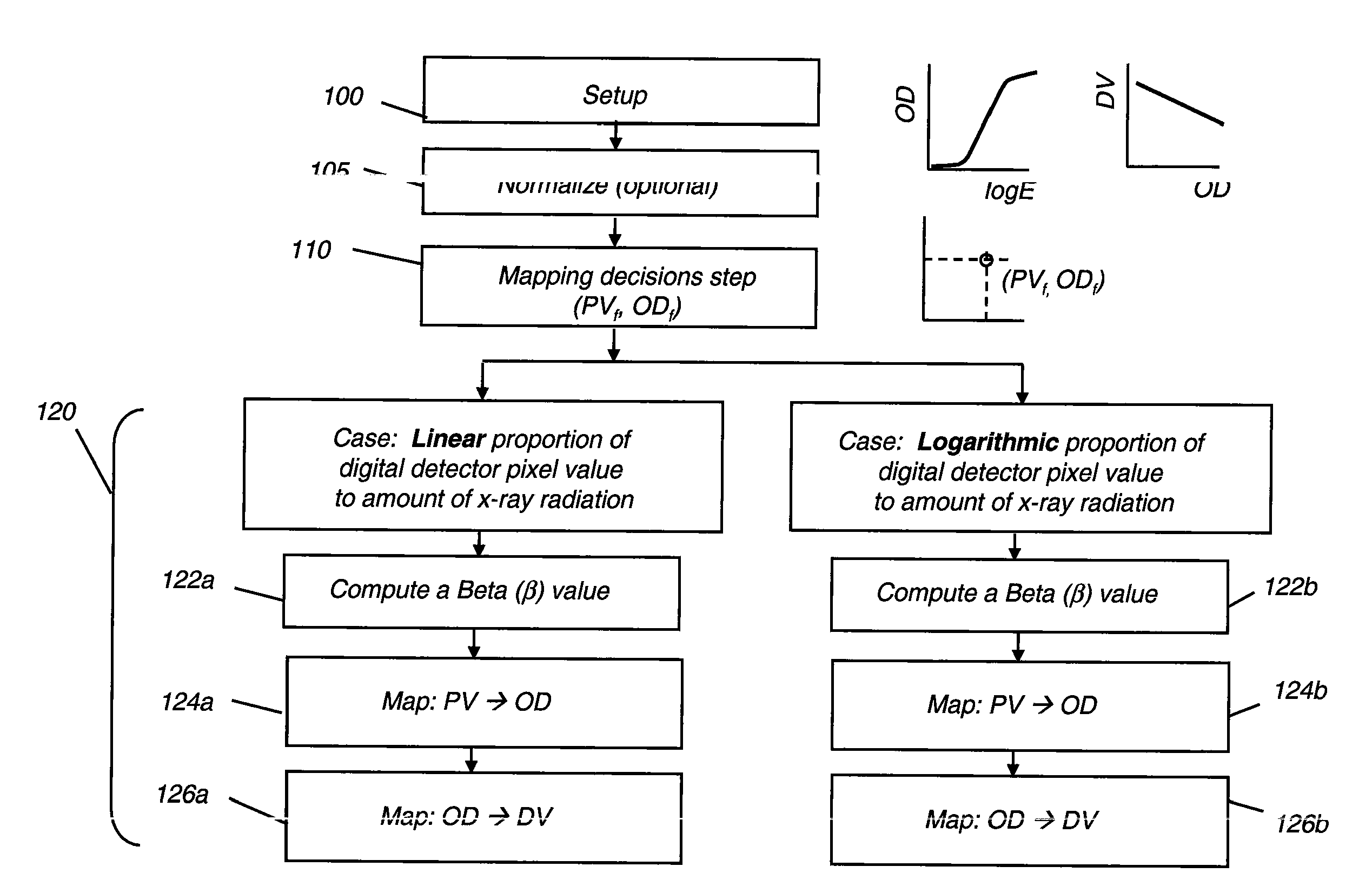
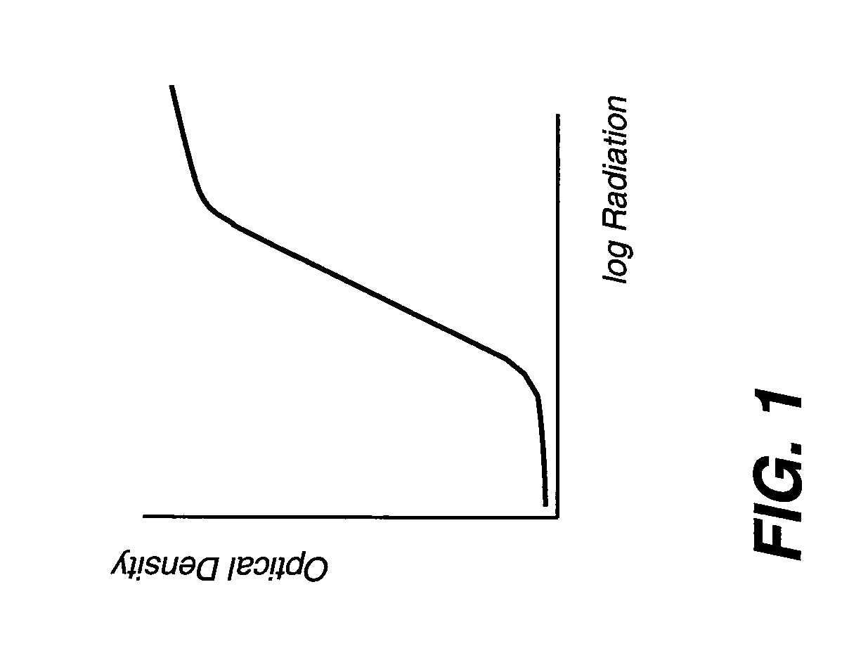
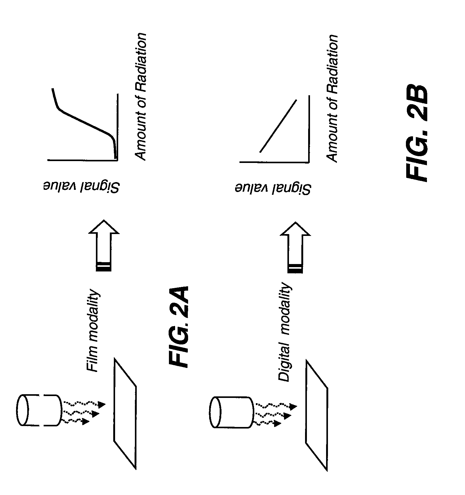
[0019]It is useful to clarify a number of terms that are used throughout the following detailed description and claims. The term “digitized data” refers to image data that originates with an image formed on a photosensitive film medium. In conventional x-ray workflow, an x-ray image is obtained on a sheet of film, is developed, and is then scanned to convert the film image into digitized data for processing and storage. The scanning process is carried out on a screened film or scanned film (SF) system. In conventional terminology, this data is said to be in digitized data space. In the context of the present disclosure, the label DV denotes values in this Digitized Value space (DV space).

[0020]In contrast, the term “digital receiver data” refers to digital data signals obtained directly from a digital receiver, such as that provided in a CR or DR system. This data is said to be in digital receiver data space. In the context of the present disclosure, the label PV denotes pixel value...


PUM


Abstract

A method for mapping radiological image data from a source imaging system to a target imaging system obtains sensitometric response data for both the source and the target imaging systems and identifies an anchor point that relates a specified source signal value to a specified target signal value. A transform is applied to map a range of source signal values to a corresponding range of target signal values, wherein the transform is defined according to the sensitometric response data obtained for both the source and the target imaging systems and according to the identified anchor point.

Description

FIELD OF THE INVENTION[0001]The invention relates generally to radiological images and more particularly to a method for correlating image data obtained from different radiographic imaging systems.BACKGROUND OF THE INVENTION[0002]For many years, conventional radiography employed photosensitive silver-halide film medium for recording images of human and animal tissue. Then, in order to take advantage of advances in digital image processing, many radiographic images originally recorded on film were digitized in order to provide these images in digital data format. This image data could then be analyzed by sophisticated imaging algorithms capable of detecting various conditions that might not be easily be discerned from the film image, even by the trained eye of a skilled diagnostician. A considerable amount of research and development effort has been expended over the last several years to develop and refine image processing algorithms that assist the diagnostician in assessing digiti...

Claims


Application Information

Patent Timeline
IPC IPC(8): G01N23/04
CPCA61B6/585A61B6/4233
Inventor YANG, CHANG-YING JOSEPHHUO, ZHIMIN
Owner CARESTREAM HEALTH INC
Why Eureka
Try Eureka