Apparatus and method for detecting a fetal heart rate

a technology of fetal heart rate and applicator, which is applied in the field of applicator and fetal heart rate detection, can solve the problems of limiting the use of systems in hospitals, limiting the use of doppler ultrasound, and invasive long-term recording of fhr using ultrasound

Inactive Publication Date: 2009-07-30
MONICA HEALTHCARE
View PDF7 Cites 20 Cited by
  • Summary
  • Abstract
  • Description
  • Claims
  • Application Information

AI Technical Summary

Benefits of technology

[0028]Furthermore, this allows a portable device to be produced which can be carried by the mother, thereby allowing the measurements to be made over a longer time interval than has previously been achieved.
[0078]By accepting that the signal can only be detected in any patient for approximately 40% of the time using a single detector, we have developed a system that is portable, can be used for 24 hours or longer, has more than one recording channel and is extremely low noise. By initially deducing the baby position a small array of electrodes can be placed around the fetus on the mother=s abdomen−thus increasing the detection rate. By recording over 24 hours using our improved technique will typically result in at least 10 hours of data. This long term, non-invasive collection of fHR data allowing mothers freedom to function in their normal environment has never been achieved before.

Problems solved by technology

Such systems are therefore limited to use in a hospital environment.
However, due to the nature of the ultrasound signal being directed at the fetus then long term recordings of fHR using ultrasound may be considered as invasive.
Although this invasive nature has not been clinically substantiated, the use of Doppler ultrasound is still limited to short time periods.
However, the system requires complex circuitry and the SQUID transducer must be cooled to liquid helium temperatures, thus resulting in these systems being both expensive and large.
Although long term recordings can be made they are nevertheless carried out in an environment, which due to the nature of the surrounding equipment, is neither comfortable nor easily accessible to a wider community.
Such a technique can be applied to the fetal heart during pregnancy but is highly susceptible to extraneous audible noises.
However, the technique is highly invasive and in some cases extremely risky, and accordingly can only be performed by a highly skilled medical team.
As a result of the system requiring separate processing apparatus to process the obtained data as it is generated, the unit is not portable.
Also, the system only uses a single channel and this means that for a large proportion of the time during which measurements are made, the fetal heart rate cannot be detected.
Not only does this therefore require that the mother spend a period of time in hospital for the monitoring to take place, but this also means that the equipment is not used for long-term fetal heart beat detection.
However, Agood@ data is infrequently obtained and typically in 60% of cases the data is Abad@ meaning that the fetal ECG (fECG) is undetectable.
Hence out of 10 mothers visiting the hospital the technique, in this form, will only be successful on typically 4 pregnancies—a figure which makes the technique unacceptable for routine spot checks of abdominal fECG.
As can be determined from the results described above, these two parameters have in the past been so disappointing that results were never considered acceptable and therefore the technique has not been routinely adopted in clinical practice.
Accordingly, it is not currently possible to record reliably a long-term fetal heart rate using a portable and non-invasive technique in the home.

Method used


Image

  • Apparatus and method for detecting a fetal heart rate
  • Apparatus and method for detecting a fetal heart rate
  • Apparatus and method for detecting a fetal heart rate

Examples


Embodiment Construction

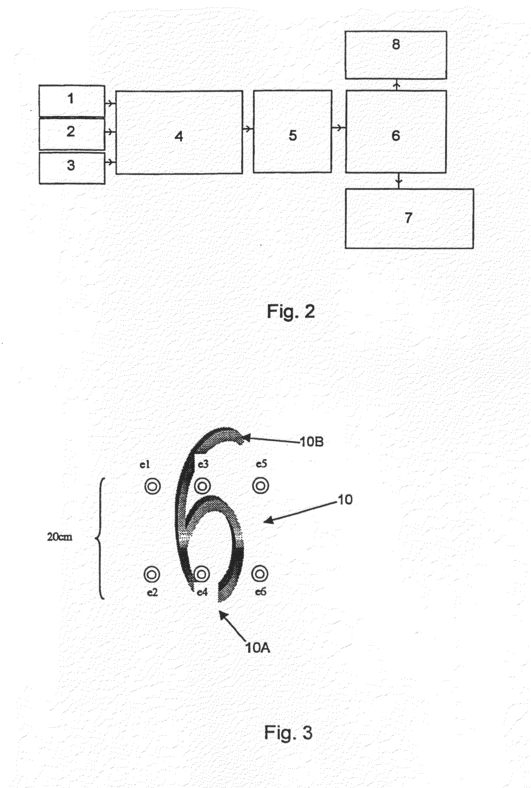
[0094]FIG. 2 is a block diagram showing apparatus for detecting the heart beat of a fetus. The apparatus comprises three detectors 1,2,3 which are coupled to an amplification and filter stage 4. The output of the amplification and filter stage 4 is coupled to an analogue-to-digital converter 5, which is in turn connected to a processor 6. The processor 6 is coupled to a memory 7 and a display 8.

[0095]The system operates as follows. Each detector 1,2,3 consists of two passive cutaneous conductive electrodes positioned on the abdomen of the mother so as to detect ECG signals generated in the region of the mother's abdomen. An example of a suitable electrode arrangement is shown in FIG. 3. In this case, the electrodes e1,e2 correspond to detector 1, electrodes e3,e4 correspond to detector 2 and electrodes e5,e6 correspond to detector 3. Reference numeral 10 represents the fetus, with 10A representing the head and 10B the fetal back.

[0096]Electrical signals detected by the detectors 1,2...


PUM


Abstract

An apparatus and method for detecting the heart rate of a fetus. The apparatus includes three detectors detecting heart beats of the fetus, each detector including at least two electrodes detecting ECG signals, the detectors being positioned on an abdomen of a mother in use. The apparatus also includes a processor coupled to the detectors, the processor being adapted to process the ECG signals received from the detectors and determine a heart rate of the fetus. The method of determining the heart rate of the fetus includes determining a position of the fetus within a womb, placing the detectors on an abdomen of the mother, monitoring the ECG signals obtained from the detectors for a predetermined length of time, and processing the ECG signals obtained from the detectors positioned on the abdomen of the mother to determine heart beats of the mother.

Description

CROSS-REFERENCE TO RELATED APPLICATIONS[0001]This application is a divisional of application Ser. No. 10 / 089,997, filed Sep. 23, 2002, allowed. This application is based upon and claims the priority of Great Britain application no. 9923955.0, filed Oct. 8, 1999, PCT application no. PCT / GB00 / 03843, filed Oct. 6, 2000 and U.S. patent application Ser. No. 10 / 089,997, filed Sep. 23, 2002, the contents being incorporated herein by reference.BACKGROUND OF THE INVENTION[0002]1. Field of the Invention[0003]The present invention relates to an apparatus and a method for detecting the heart rate of a fetus.[0004]2. Description of the Related Art[0005]It is useful to be able to detect the heart beat of fetuses as this can provide information regarding the health of the fetus during the progress of pregnancy. Currently there are four main methods for detecting fetal heart rates during pregnancy and these involve the use of Doppler ultrasound, a SQUID magnetometer, phonocardiography, and abdomina...

Claims


Application Information

Patent Timeline
Patent Type & Authority Applications(United States)
IPC IPC(8): A61B5/0444A61B5/024A61B5/344
CPCA61B5/02411A61B5/4362A61B5/0444A61B5/344
Inventor HAYES-GILL, BARRIEJAMESCROWE, JOHNPIERI, JEAN-FRANCOIS
Owner MONICA HEALTHCARE
Why Eureka
Try Eureka