Stent delivery under direct visualization

Active Publication Date: 2009-08-13
INTUITIVE SURGICAL OPERATIONS INC
View PDF105 Cites 221 Cited by
  • Summary
  • Abstract
  • Description
  • Claims
  • Application Information

AI Technical Summary

Benefits of technology

[0024]In yet another variation, a deployment catheter may utilize a hood having an angled interface which is angled relative to the catheter. The asymmetric slanted hood may be used to facilitate navigation within the

Problems solved by technology

However, such imaging balloons have many inherent disadvantages.
For instance, such balloons generally require that the balloon be inflated to a relatively large size which may undesirably displace surrounding tissue and interfere with fine positioning of the imaging system against the tissue.
Moreover, the working area created by such inflatable balloons are generally cramped and limited in size.
Furthermore, inflated balloons may be susceptible to pressure changes in the surrounding fluid.
For example, if the environment surrounding the inflated balloon undergoes pressure changes, e.g., during systolic and diastolic pressure cycles in a beating heart, the constant pressure change may affect the inflated balloon volume and its positioning to produce unsteady or undesirable conditions for optimal tissue imaging.
Additionally, imaging balloons are subject to producing poor or blurred tissue images if the balloon is not firmly pressed against the tissue surface because of intervening blood between the balloon and tissue.
Accordingly, these types of imaging modalities are generally unable to provide desirable images useful for sufficient diagnosis and therapy of the endoluminal structure, due in part to factors such as dynamic forces generated by the natural movement of the heart.
Moreover, anatomic structures within the body can occlude or obstruct the image acquisition process.
Also, th

Method used


Image

  • Stent delivery under direct visualization
  • Stent delivery under direct visualization
  • Stent delivery under direct visualization

Examples


Embodiment Construction

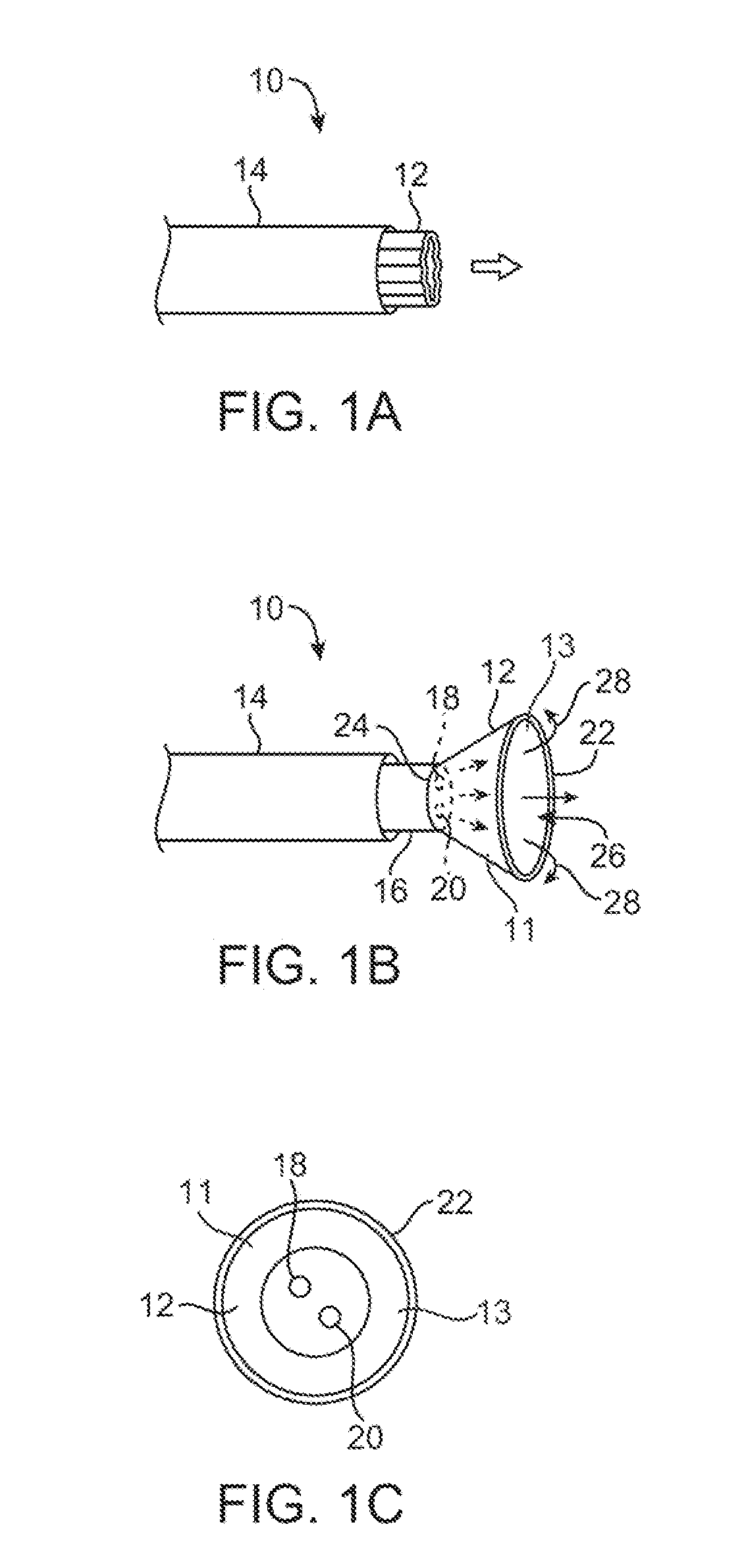
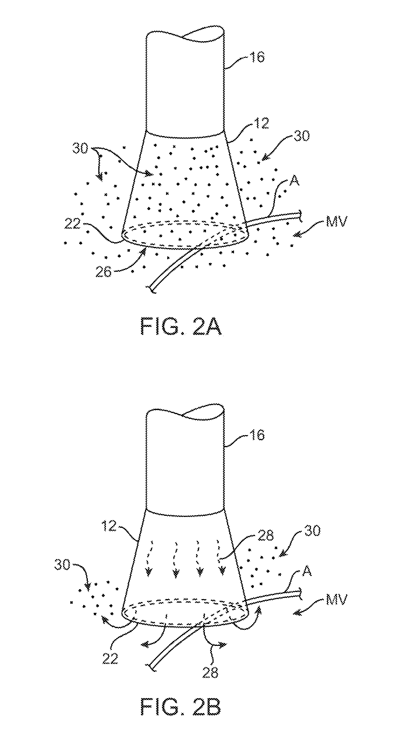
[0040]A tissue-imaging and manipulation apparatus described herein is able to provide real-time images in vivo of tissue regions within a body lumen such as a heart, which is filled with blood flowing dynamically therethrough and is also able to provide intravascular tools and instruments for performing various procedures upon the imaged tissue regions. Such an apparatus may be utilized for many procedures, e.g., facilitating transseptal access to the left atrium, cannulating the coronary sinus, diagnosis of valve regurgitation / stenosis, valvuloplasty, atrial appendage closure, arrhythmogenic focus ablation, among other procedures.

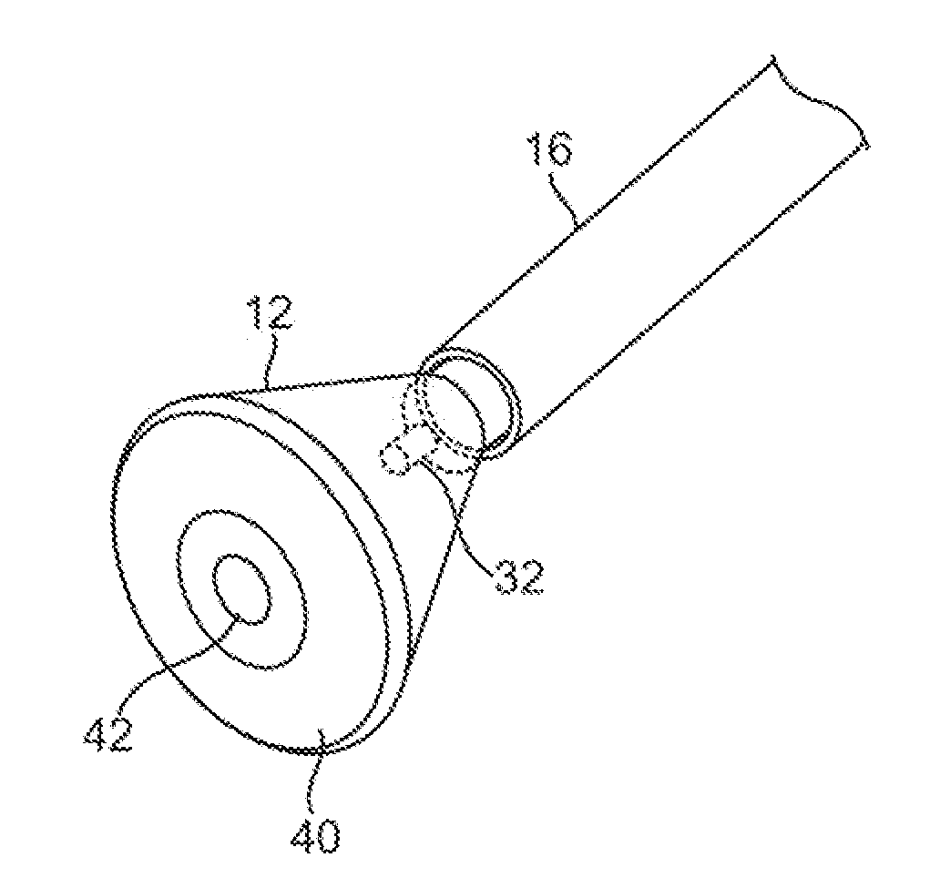
[0041]One variation of a tissue access and imaging apparatus is shown in the detail perspective views of FIGS. 1A to 1C. As shown in FIG. 1A, tissue imaging and manipulation assembly 10 may be delivered intravascularly through the patient's body in a low-profile configuration via a delivery catheter or sheath 14. In the case of treating tissue, it is gener...


PUM


Abstract

Stent delivery under direct visualization utilizing an imaging hood is described herein. A stent may be delivered and placed in or around lesions, e.g., ostial lesions, through a delivery catheter while being directly visualized via an imaging hood. A pre-delivery assessment probe may also be advanced to the desired site for accurate placement of the stent.

Description

CROSS-REFERENCE TO RELATED APPLICATIONS[0001]This application claims the benefit of priority to U.S. Prov. Pat. App. 61 / 026,795 filed Feb. 7, 2008, which is incorporated herein by reference in its entirety.FIELD OF THE INVENTION[0002]The present invention relates generally to stent delivery systems which may be used to place one or more stents along a lesion. More particularly, the present invention relates to methods and apparatus for the delivery of one or more stents along a lesion, such as an ostial lesion or along a stenosed region (e.g., caused by atherosclerotic plaque) at various locations, for instance along the coronary vessels, renal arteries, etc., while directly visualizing the tissue region.BACKGROUND OF THE INVENTION[0003]Conventional devices for visualizing interior regions of a body lumen are known. For example, ultrasound devices have been used to produce images from within a body in vivo. Ultrasound has been used both with and without contrast agents, which typica...

Claims


Application Information

Patent Timeline
IPC IPC(8): A61F2/84A61B1/04A61F2/82
CPCA61F2/958A61F2002/821A61B19/5244A61B1/04A61F2250/0039A61B34/20A61B1/00089A61B1/12A61B1/044
Inventor MILLER, DAVIDSAADAT, VAHIDPEH, RUEY-FENG
Owner INTUITIVE SURGICAL OPERATIONS INC
Why Eureka
Try Eureka