Ultrasound-imaging systems and methods for a user-guided three-dimensional volume-scan sequence

a three-dimensional ultrasound and ultrasound imaging technology, applied in tomography, applications, instruments, etc., can solve the problems of affecting the frame rate of real-time imaging systems, unable to provide provisions to permit the technician to reduce the size of volumes, and a large amount of time to acquire large volumes, so as to influence the rate of target-volume acquisition and optimize frame rates

Inactive Publication Date: 2009-08-13
KONINKLIJKE PHILIPS ELECTRONICS NV
View PDF8 Cites 29 Cited by
  • Summary
  • Abstract
  • Description
  • Claims
  • Application Information

AI Technical Summary

Benefits of technology

[0020]The present invention provides an ultrasound-imaging system and method for acquiring a user-identified target volume in a time-efficient manner. An operator interacting with the ultrasound-imaging system identifies the volumetric size, the location, the view angle, etc. of a target volume. In response to the operator input, the ultrasound-imaging system may alter a scan sequence, an acoustic-line direction, and an acoustic-line spacing to acquire a three-dimensional image in a time-efficient manner. The operator can significantly

Problems solved by technology

As a result, no provisions were provided to permit the technician to reduce the size of a volume-under-observation (VUO).
However, it takes a considerable amount of time to acquire a large volume scan, which negatively impacts the frame rate in a real-time imaging system.
In non-real-time systems, it is the total time of acquisition that is negatively impacted.
However, this approach significantly increases the cost and the size of the resulting ultrasound-imaging system.
This 16x parallel beamformer architecture is undesirable as the realizable three-dimensional resolution comes at a significant cost, especially when compared with prior art two-dimensional imaging systems.
First, the cost for each beamformer makes the prior art three-dim

Method used


Image

  • Ultrasound-imaging systems and methods for a user-guided three-dimensional volume-scan sequence
  • Ultrasound-imaging systems and methods for a user-guided three-dimensional volume-scan sequence
  • Ultrasound-imaging systems and methods for a user-guided three-dimensional volume-scan sequence

Examples


Embodiment Construction

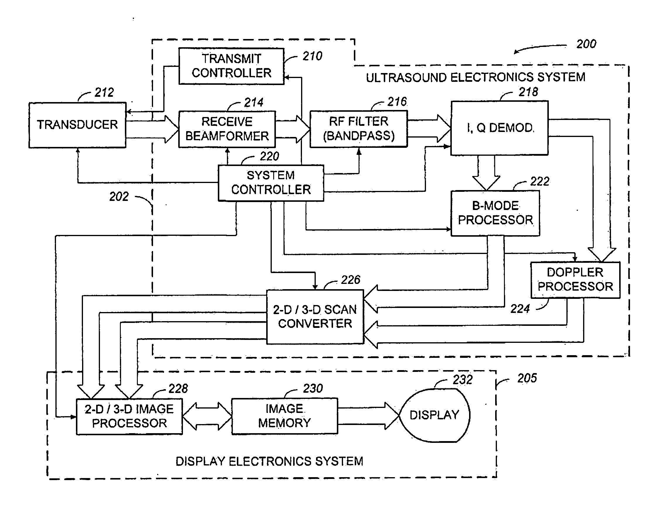
[0037]The three-dimensional ultrasound-imaging system and method of the present invention will now be specifically described in detail in the context of an ultrasound-imaging system that creates and displays brightness-mode (B-Mode) images, or gray-scale images, as well as, color-flow or Doppler-mode images which are well known. However, it should be noted that the teachings consistent with the improved three-dimensional ultrasound-imaging system and method of the present invention may be practiced using other ultrasound-imaging systems that are suited for the method, as will be apparent to those skilled in the art.

System Architecture and Operation

[0038]An exemplar architecture of an embodiment of an ultrasound-imaging system capable of implementing the method of the present invention is illustrated by way of a functional block diagram in FIG. 4 and is generally denoted by reference numeral 200. Note that many of the functional blocks illustrated in FIG. 4 define a logical function ...


PUM


Abstract

An ultrasound-imaging system and method is provided that permits an operator to acquire an image of a volume-of-interest in a time critical fashion, that is capable of referencing the volume rendering to a standard two-dimensional imaging mode, and permits the operator to selectively choose a number of display-mode parameters that result in an operator directed view of the volume-of-interest. The ultrasound-imaging system comprises an input device configured to receive a plurality of imaging parameters and a controller in communication with the input device. The ultrasound-imaging system generates an operator-directed transmit-beam scan sequence in response to the imaging parameters and transmits a spatially modified transmit-beam scan sequence over a portion of the volume-scan range of the ultrasound-imaging system. Moreover, the ultrasound-imaging system provides the flexibility for an operator to direct a plurality of operator-configurable multi-dimensional views.

Description

FIELD OF THE INVENTION[0001]The present invention generally relates to three-dimensional ultrasound-imaging systems for the purpose of medical diagnosis, and more particularly, to an improved three-dimensional ultrasound-imaging system and method that provides an increased frame rate or an optimized image-acquisition time.BACKGROUND OF THE INVENTION[0002]Ultrasonic imaging has become an important and popular diagnostic tool with a wide range of applications. Particularly, due to its non-invasive and typically non-destructive nature, ultrasound imaging has been used extensively in the medical profession. Modern high-performance ultrasound-imaging systems and techniques are commonly used to produce both two-dimensional and three-dimensional diagnostic images of internal features of an object, (e.g., portions of the anatomy of a human patient). A diagnostic ultrasound-imaging system generally uses a wide-bandwidth transducer to emit and receive ultrasound signals. The ultrasound-imagin...

Claims


Application Information

Patent Timeline
IPC IPC(8): A61B8/14G01S7/52G01S15/89
CPCA61B8/467A61B8/483G01S15/8993G01S15/8925G01S7/52068A61B8/466A61B8/469
Inventor THIELE, KARL ERHARDSOLOMON, RODNEY J.ADLEMAN, GEORGESAVORD, BERNARD
Owner KONINKLIJKE PHILIPS ELECTRONICS NV
Why Eureka
Try Eureka