Amnion-derived stem cells and uses thereof
- Summary
- Abstract
- Description
- Claims
- Application Information
AI Technical Summary
Benefits of technology
Problems solved by technology
Method used
Image
Examples
example 1
[0112]ADSC Isolation, Culture and Cloning
[0113]Rat amnion membrane is mechanically separated from the chorion of embryonic day 18.5 (E18.5) Sprague-Dawley rat embryos. The tissue is washed extensively with phosphate buffered saline (PBS) and subsequently cut into small pieces. Membrane fragments are placed in 6-well plastic tissue culture dishes in a minimal volume (0.5 ml) of Dulbecco's modified Eagle's medium [DMEM; Invitrogen, Carlsbad, Calif.] supplemented with 20% Fetal Bovine Serum [FBS; Atlanta Biologicals, Atlanta, Ga.] to encourage attachment. Cells begin to emerge from the explanted tissues within 24 hours of plating. After one week the tissue explants are removed and the remaining adherent cells are trypsinized and re-plated into 100 mm plastic culture dishes. Cultures are passaged at confluency. All cultures are maintained under a humidified atmosphere of 5% CO2 at 37° C. ADSCs used in these studies are extensively propagated up to 50 passages in vitro.
[0114]Generation o...
example 2
rived Stem Cell Isolation and Characterization
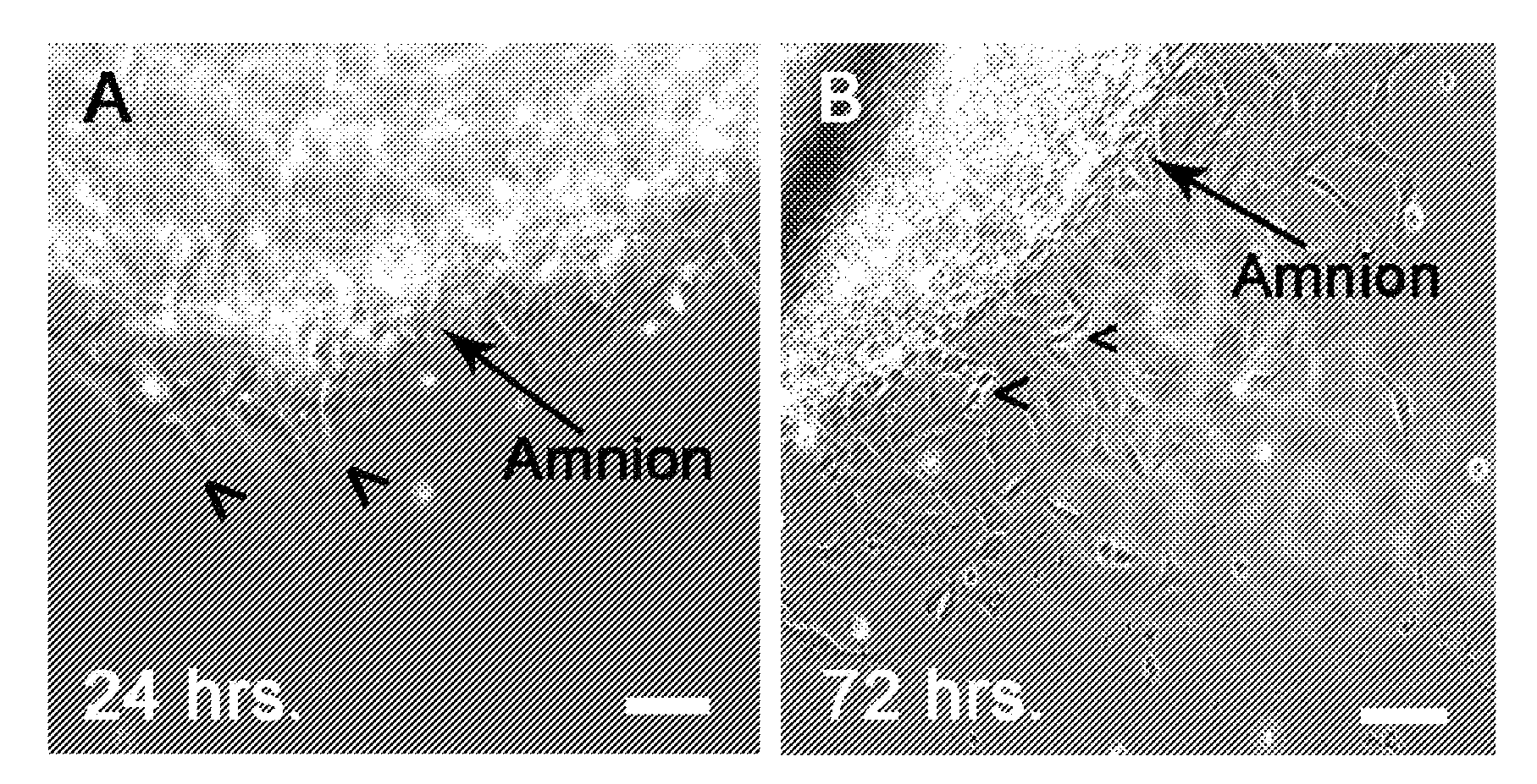
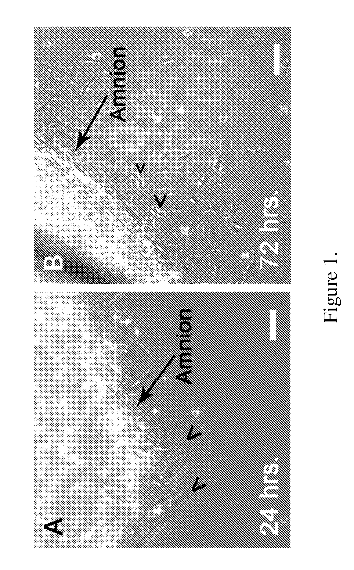
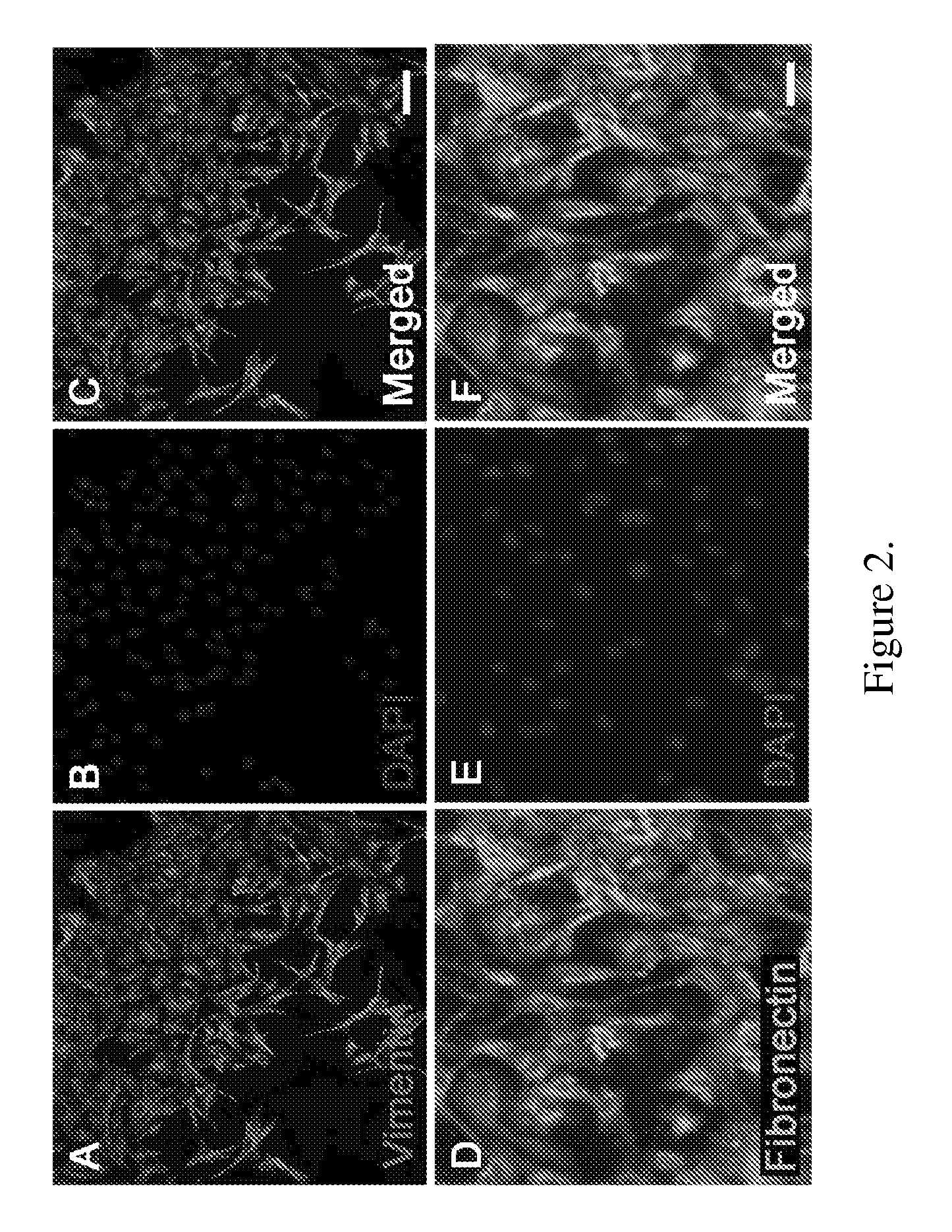
[0142]Amniotic membranes are isolated from embryonic day 18.5 (E18.5) rats. To avoid contamination with previously identified fetal stem cell populations residing in the rat placenta or umbilical cord, amnion tissue is isolated from the dorsal part of the amniotic sac. The amnion is mechanically peeled from the chorion and subsequently cut into small pieces and placed into tissue culture. Within 24 hours, cells begin to emerge from the tissue explants (FIG. 1A). By 72 hours numerous ameboid shaped cells migrate from the tissue pieces. Cell doublets are clearly visible, indicating cell proliferation (FIG. 1B). Immunocytochemical analysis of primary cultures reveal that by 24 hours fibronectin-positive (+) cells are visible at the edge of the tissue explants. In contrast, cytokeratin-19 (CK-19)+ epithelial cell remain within the tissue pieces and do not migrate from the explants. At 72 hours close to 100% of the migrating cells are vimenti...
example 3
fferentiation of ADSCs
[0150]To examine neuroectodermal potential, ADSCs are exposed to a defined neural induction medium (NIM). ADSCs responds slowly to the induction media assuming elongated fibroblastic morphologies within 24 hours (FIGS. 5, A and B). Over the next several days, greater than 75% of the cells convert from flat, ameboid figures to cells displaying compact, light refractile cell bodies (FIG. 5C). ADSCs elaborate long processes (FIG. 5C) some extending more than 500 μm from the cell body. In many cases, the cellular processes form networks (FIG. 5D) with neighboring cells similar to those found in primary neural cultures.
[0151]Morphological changes of differentiated ADSCs are accompanied by consistent changes in gene expression. ADSCs maintain the expression of some neuronal genes such as NF-M, and up-regulate other neural genes (FIG. 5, E-H). For instance, tau, a microtubule associated protein found in mature neurons, is expressed only in NIM-treated cultures (FIG. 5...
PUM
 Abstract
Description
Claims
Application Information
 - R&D
- Intellectual Property
- Life Sciences
- Materials
- Tech Scout
- Unparalleled Data Quality
- Higher Quality Content
- 60% Fewer Hallucinations
Browse by: Latest US Patents, China's latest patents, Technical Efficacy Thesaurus, Application Domain, Technology Topic, Popular Technical Reports.
© 2025 PatSnap. All rights reserved.Legal|Privacy policy|Modern Slavery Act Transparency Statement|Sitemap|About US| Contact US: help@patsnap.com


