Method for detecting mammary cancer cells
a mammary cancer and cell technology, applied in the field of mammary cancer cell detection, can solve the problems of long time for examination completion, inaccurate data, unsatisfactory methods, etc., and achieve the effect of rapid and simple detection and rapid cure of mammary cancer
- Summary
- Abstract
- Description
- Claims
- Application Information
AI Technical Summary
Benefits of technology
Problems solved by technology
Method used
Image
Examples
example 1
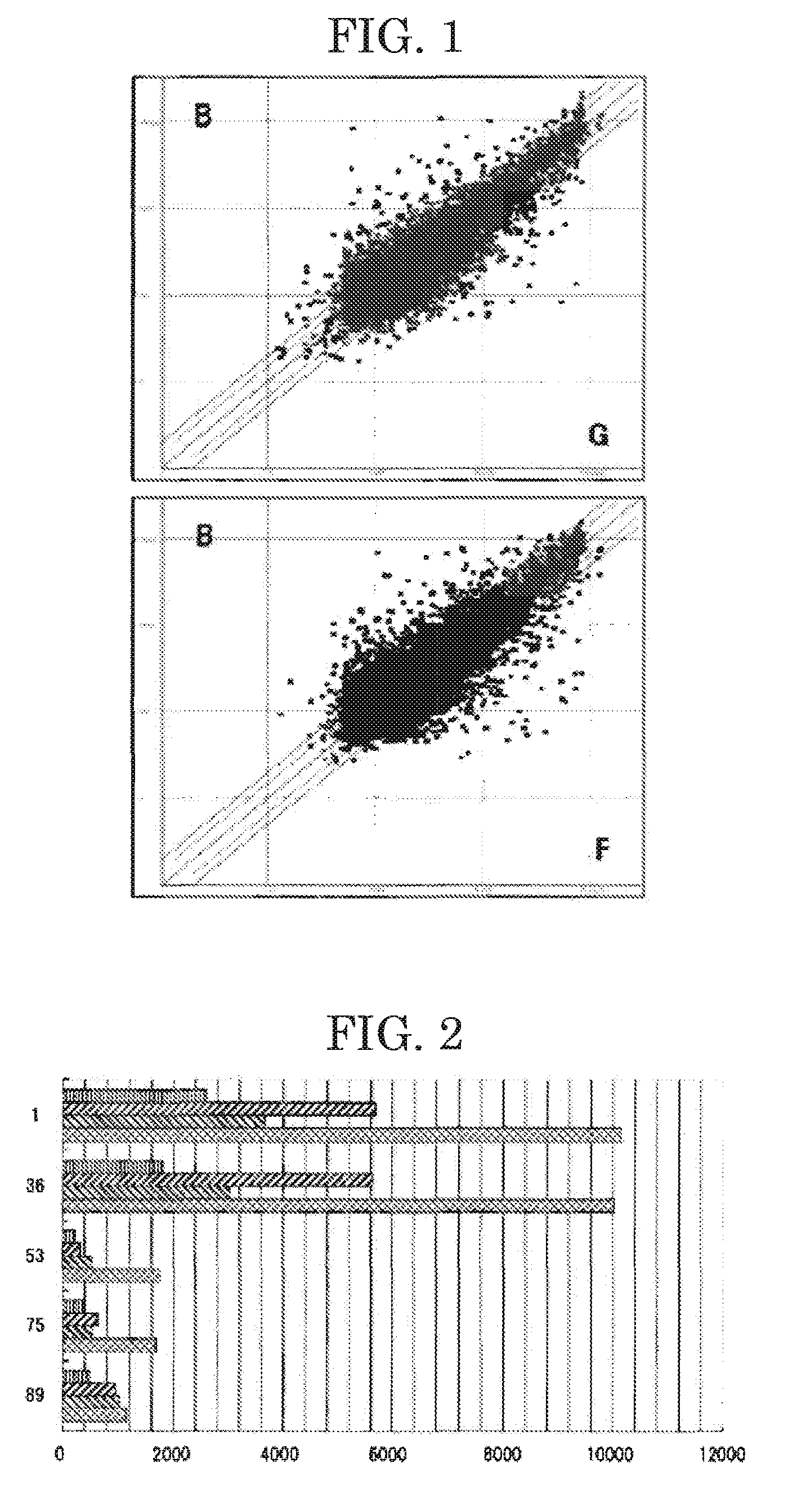
[0027]The present invention will next be described by way of Examples, which should not be construed as limiting the present invention thereto. In the Examples, human mammary cancer tissue and normal mammary gland tissue were analyzed for gene expression level (RNA transcription level) through high coverage expression profiling analysis (HiCEP technique) (Nucleic Acids Res., 2003, vol. 31(16), p. 94)—one known technique for gene expression analysis. And, the mammary cancer tissue was compared with the normal mammary gland tissue in terms of expression levels of the genes thereof.
[0028]Specifically, one normal mammary gland tissue (sample #N) and three human mammary cancer tissues (samples #B, #F and #G) (Table 1) were treated using an RNeasy kit (product of QUIAGEN Co.) with a routine method for the kit, to thereby extract total RNA samples therefrom. Then, using a Super Script First Strand Synthesis System for RT-PCR (product of Invitrogen Corporation), reverse transcription was ca...
example 2
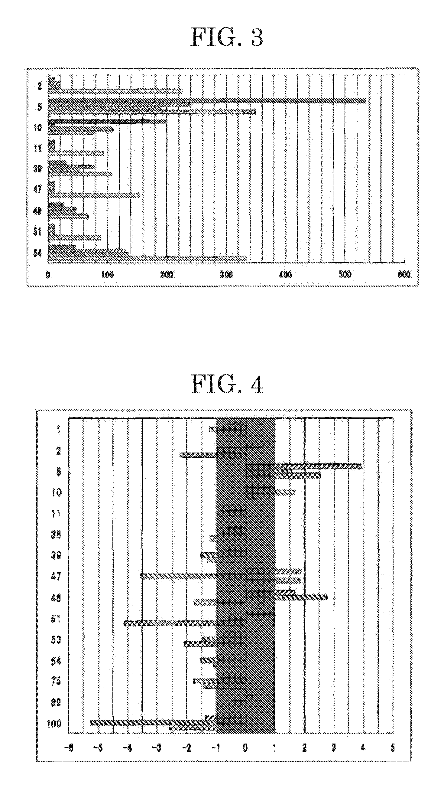
[0031]In this Example, quantitative RT-PCR was performed on the 14 genes, whose expression levels had been found to be different from those of the genes of the normal mammary gland tissue sample in Example 1, and their expression levels in the mammary cancer tissue samples were compared with those in the normal mammary gland tissue sample. Specifically, total RNA samples were extracted from the tissue samples with a routine method. Then, reverse transcription was performed using as a template each total RNA sample (1 μg), to thereby synthesize single-stranded cDNA fragments complementary to mRNA fragments contained in the total RNA sample. Subsequently, using the thus-synthesized single-stranded cDNA fragment serving as a template and SYBER Green PCR Master (product of TOYOBO CO., LTD.), real-time PCR was performed through a routine method with a real-time PCR device (Light Cycler, product of Roche Diagnostics K.K.). On the basis of the obtained gene amplification curve, the genes o...
example 3
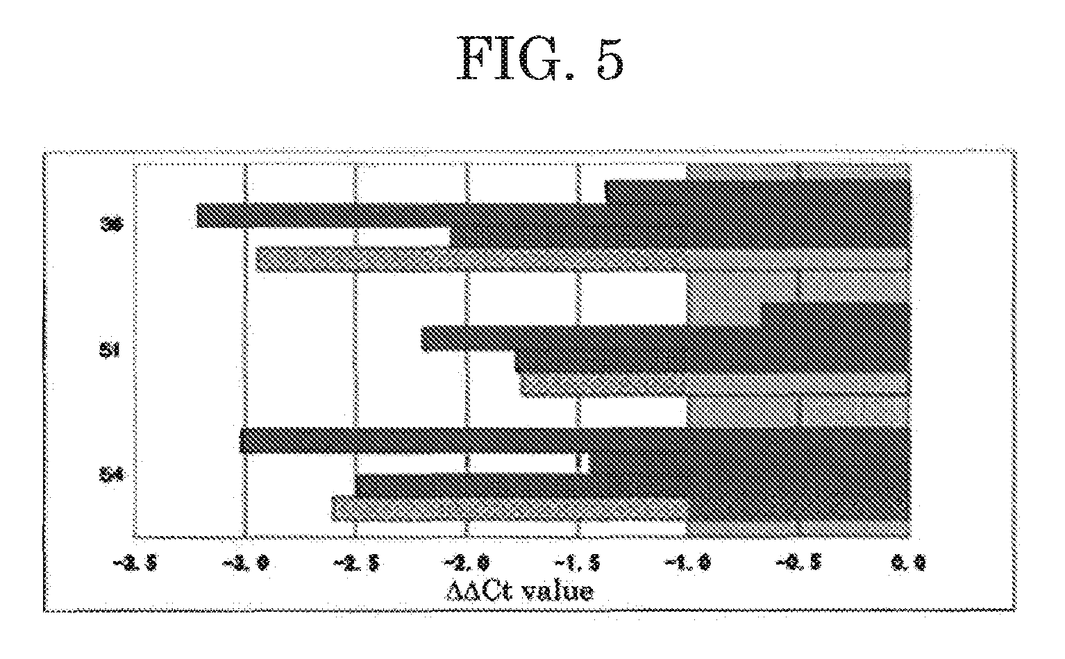
[0033]In this Example, quantitative RT-PCR was performed on three genes of the 11 genes, whose expression levels had been found to be particularly different from those of the genes of the normal mammary gland tissue sample in Example 2, and their expression levels in mammary cancer tissue samples were compared with those in the normal mammary gland tissue sample of Example 2. The mammary cancer tissue samples used were mammary cancer tissue samples #D, #E, #H and #I (Table 3) different from those used in Example 2. Specifically, total RNA samples were extracted from the tissue samples with a routine method. Then, reverse transcription was performed using as a template each total RNA sample (1 μg), to thereby synthesize single-stranded cDNA fragments complementary to mRNA fragments contained in the total RNA sample. Subsequently, using the thus-synthesized single-stranded cDNA fragment serving as a template and SYBER Green PCR Master (product of TOYOBO CO., LTD.), real-time PCR was p...
PUM
| Property | Measurement | Unit |
|---|---|---|
| diameter | aaaaa | aaaaa |
| of time | aaaaa | aaaaa |
| capillary electrophoresis | aaaaa | aaaaa |
Abstract
Description
Claims
Application Information
 - R&D
- Intellectual Property
- Life Sciences
- Materials
- Tech Scout
- Unparalleled Data Quality
- Higher Quality Content
- 60% Fewer Hallucinations
Browse by: Latest US Patents, China's latest patents, Technical Efficacy Thesaurus, Application Domain, Technology Topic, Popular Technical Reports.
© 2025 PatSnap. All rights reserved.Legal|Privacy policy|Modern Slavery Act Transparency Statement|Sitemap|About US| Contact US: help@patsnap.com


