Optical detection of seizure, a pre-seizure state, and cerebral edema and optical fiber detection of the same

a seizure and seizure state technology, applied in the field of neural seizure detection and brain tissue edema detection methods, can solve the problem that scattering changes cannot be detected at small source-to-detector separation, and achieve the effect of reducing the degree of optical scattering by neural brain tissu

Inactive Publication Date: 2009-12-17
RGT UNIV OF CALIFORNIA
View PDF5 Cites 11 Cited by
  • Summary
  • Abstract
  • Description
  • Claims
  • Application Information

AI Technical Summary

Benefits of technology

[0013]We have discovered particular optical parameters which are useful in detection of the seizure and pre-seizure state in the brain. Specifically, we have demonstrated that the degree of optical scattering by neural brain tissue decreases before seizures are detected with electroencephalography (EEG), and before becoming manifested clinically. The optical detection of scattering changes allows us to build an apparatus which can predict and record seizures before they occur and trigger interventions to prevent or abort them. Optical apparatus capable of detecting these scattering changes can also be used to image and map regions of brain tissue undergoing seizure. The configuration of our apparatus allows reliable detection of the pre-seizure state earlier than any previously described system, either optical or electrographic.
[0016]Seizure detection through optical fibers provides better signal to noise and less susceptibility to interference and movement artifact than EEG. It allows for optical signals to be obtained from anywhere in the brain into which fibers may be inserted, while current optical systems are limited to within a few millimeters of the cortical surface.
[0018]There is currently no method to predict seizure onset using optical techniques. Prediction of seizure onset provides clinical opportunities for therapeutic interventions for prevention or early arrest of clinical seizures. Seizure detection through measurement of optical scattering provides a reliable method of seizure prediction which has not hitherto been available. This process can be utilized by a variety of optical devices which are capable of measuring scattering coefficients, well known to those with ordinary skill in the art, and is not limited to the illustrated embodiment. The detection of the pre-seizure state has potential uses for seizure early warning systems, closed-loop seizure termination and prevention devices, improved intra-operative mapping of epileptic foci, and optical recording of seizure activity.
[0020]This methodology or apparatus is also adaptable to a clinically functional implantable probe for direct measurement of cerebral edema in a variety of neurological and neurosurgical conditions. It can provide early warning of pathologic brain swelling well before the currently measurable late sequelae of increased intracranial pressure or hemodynamic changes.
[0022]Direct detection of cerebral edema allows measurement of one of the primary physiologic events in the cascade of neurologic deterioration from a variety of causes. Earlier detection could allow potentially lifesaving intervention sooner than current monitoring equipment allows. An optical fiber edema probe could be incorporated into a variety of commonly used or conventional intracranial monitoring devices, including pressure transducers, ventricular drainage catheters, tissue oxygenation probes, or inserted as a standalone device into an area of interest. The scattering and absorption related changes would then be analyzed to provide an edema index on a continuous basis to aid physicians in clinical decision making.
[0024]In one embodiment the physiological event is a seizure and the method further comprises the step of mediating the neural activity of the neural tissue before onset of a seizure upon a determination of the signature temporal pattern, such as preventing or reducing symptoms of the seizure.

Problems solved by technology

However scattering changes can only be detected at small source-to-detector separations.

Method used


Image

  • Optical detection of seizure, a pre-seizure state, and cerebral edema and optical fiber detection of the same
  • Optical detection of seizure, a pre-seizure state, and cerebral edema and optical fiber detection of the same
  • Optical detection of seizure, a pre-seizure state, and cerebral edema and optical fiber detection of the same

Examples


Embodiment Construction

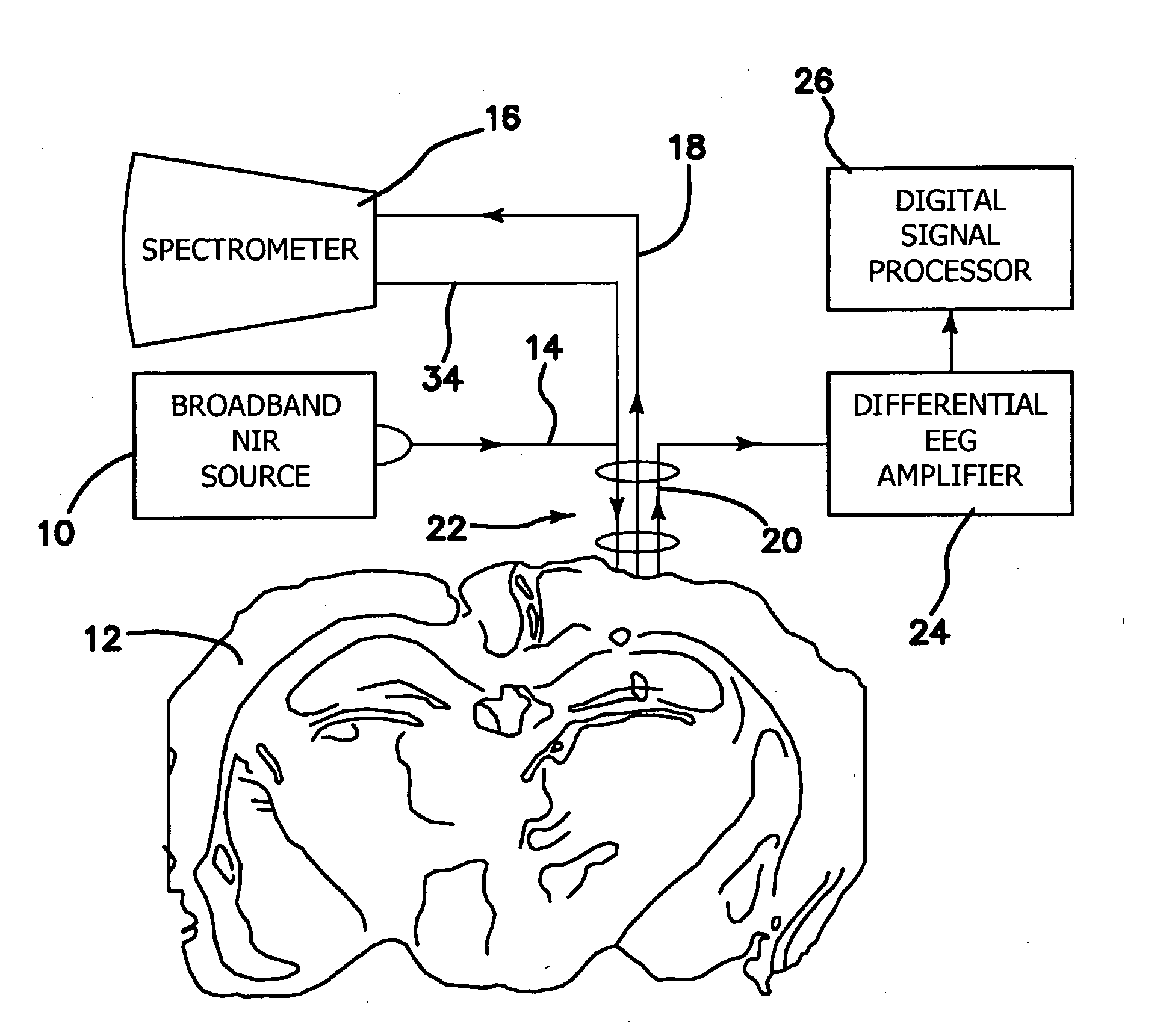
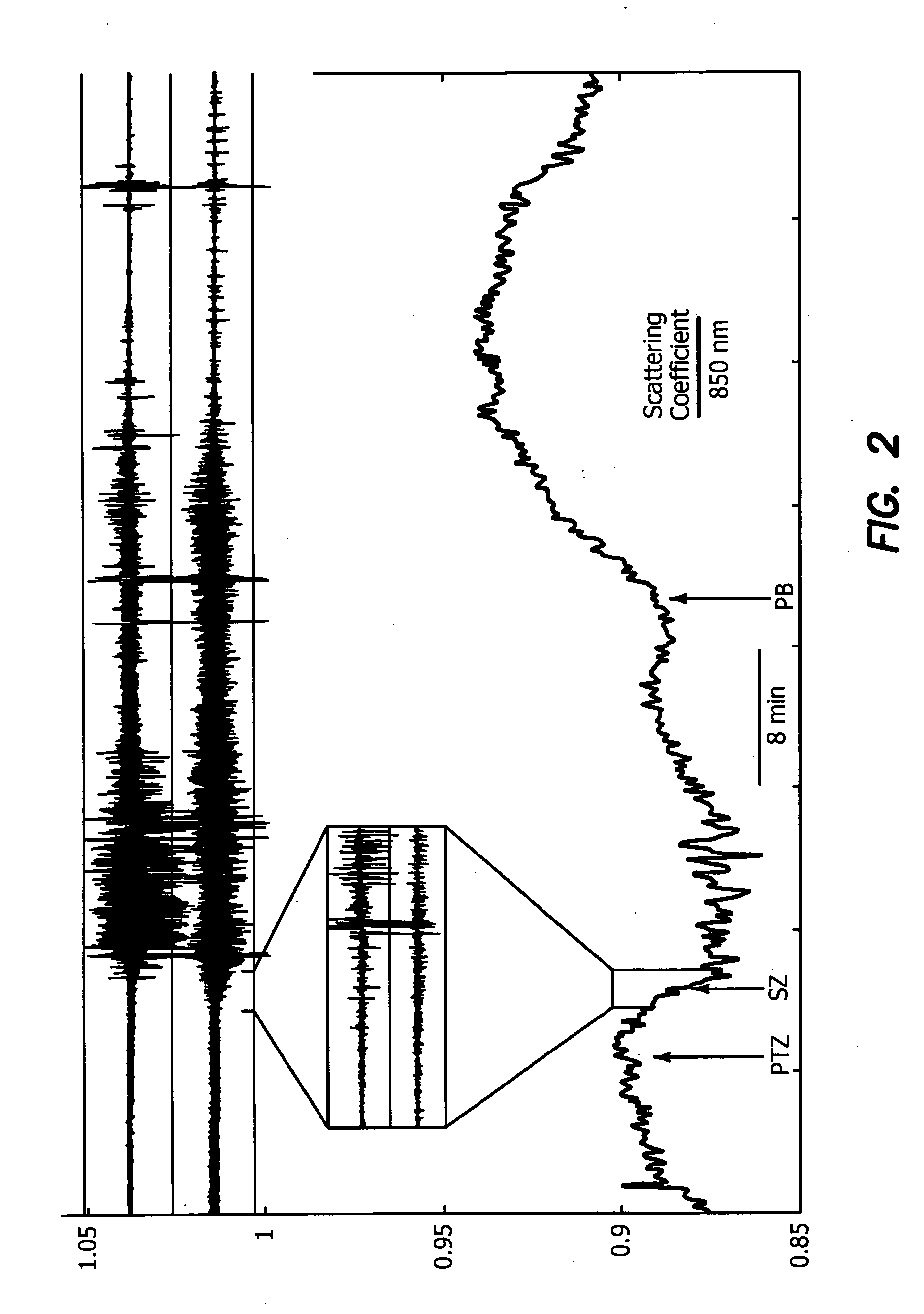
[0049]The apparatus of the illustrated embodiment measures diffuse reflectance, which at close source-detector fiber separations has been shown to correlate strongly with changes in the scattering coefficient of the brain tissue 12. This is the first study to describe the individual contribution of light scattering to the optical signal change before and during seizure activity. Our findings provide proof of principle for optical detection of a pre-seizure state on a clinically relevant timescale.

[0050]The illustrated embodiment of the invention in a laboratory demonstration as diagrammatically depicted in FIG. 4 utilizes a source 10, which illuminates the brain tissue 12 with either broadband or specific wavelengths of radiation in the visible, near-infrared, and / or infrared region, through a delivery optic fiber 14 and a detector 16 through an optic fiber 18 to measure changes in signal intensity associated with seizure or pre-seizure activity. The source 10 and detector 16 are co...


PUM


Abstract

A method for using optical parameters to monitor for a physiological event and / or a state prior to the physiological event includes the steps of: illuminating neural tissue with diagnostic light of a predetermined frequency at a predetermined location; detecting magnitude of optical scattering by neural tissue of the diagnostic light as a function of time; and determining a signature signal of the optical scattering of the diagnostic light before the physiological event in the neural tissue becomes clinically manifested. An apparatus includes a source of diagnostic light of a predetermined frequency for illuminating neural tissue at a predetermined location, a detector of optical scattering and / or optical absorption by neural tissue of the diagnostic light as a function of time, and a signal processor for determining a signature signal of the optical scattering and / or optical absorption of the diagnostic light before the physiological event becomes clinically manifested.

Description

RELATED APPLICATIONS[0001]The present application is related to U.S. Provisional Patent Application Ser. No. 60 / 972,136, filed on Sep. 13, 2007, which is incorporated herein by reference and to which priority is claimed pursuant to 35 USC 119.BACKGROUND OF THE INVENTION[0002]1. Field of the Invention[0003]The invention relates to methods of detecting neural seizures and brain tissue edema and apparatus used for the same.[0004]2. Description of the Prior Art[0005]Intrinsic optical imaging (IOS) has been used for decades to map neuronal activity and seizures in cerebral cortex. It is limited insofar as it detects only changes in diffuse reflectance which primarily arise from blood perfusion changes. Further, perfusion change occurs as a result of seizure activity and thus occurs after seizure onset. Our data indicate that scattering changes, by comparison, are associated with cellular changes which begin before a seizure occurs. This is supported by previous work by Binder et. al. in ...

Claims


Application Information

Patent Timeline
Patent Type & Authority Applications(United States)
IPC IPC(8): A61B6/00
CPCA61B5/0059A61B5/0086A61B5/6848A61B5/4878A61B5/6814A61B5/4094A61B5/0042
Inventor BINDER, DEVINOWEN, CHRISTOPHERMATHEWS, MARLON
Owner RGT UNIV OF CALIFORNIA
Why Eureka
Try Eureka