Gamma guided stereotactic localization system

a stereotactic localization and gamma technology, applied in the field of suspected cancer imaging, can solve the problems of difficult to identify cancerous lesions using mammography, high false positive rate, and limited specificity, and achieve the effect of accurate positioning and suppor

Inactive Publication Date: 2010-01-21
DILON TECH
View PDF8 Cites 15 Cited by
  • Summary
  • Abstract
  • Description
  • Claims
  • Application Information

AI Technical Summary

Benefits of technology

[0007]The invention is a stereotactic gamma-guided localization technique for imaging a suspected cancer and guiding a physician in the removal of tissue samples for biopsy. The gamma-guided localization technique includes a three step procedure including localization, correlation, and verification. The localization system includes a gamma camera with a set of slant-hole collimators for producing stereo images of a region of interest. A positioning system including a fiducial marker is placed adjacent to the object to be imaged and held rigidly in place. The positioning system provides correlation of the location of the region of interest relative to the fiducial marker. A gamma emitting marker is then positioned at the calculated location of the region of interest and imaged to verify that that the calculated position corresponds to the actual location. The positioning system can then be used to accurately position and support any other hardware that needs to be positioned at the region of interest, by measurements from the fiducial marker.

Problems solved by technology

It can be difficult to identify cancerous lesions using mammography, especially when patients have dense breast tissue, multiple suspicious lesions or clusters of microcalcifications, palpable lesions not detected by mammography or ultrasound, post-surgical or post-therapeutic mass, implants, or have been taking Hormone Replacement Therapy.
It is limited, however, by its highly variable specificity, which can range from below 37% to 97%.
Combined with its high sensitivity, it is expensive, may require multiple days to complete, and produces a high false positive rate.
However, ultrasound demonstrates a low specificity rate that can produce misleading results and indicate biopsy where one may not be needed.

Method used


Image

  • Gamma guided stereotactic localization system
  • Gamma guided stereotactic localization system
  • Gamma guided stereotactic localization system

Examples


Embodiment Construction

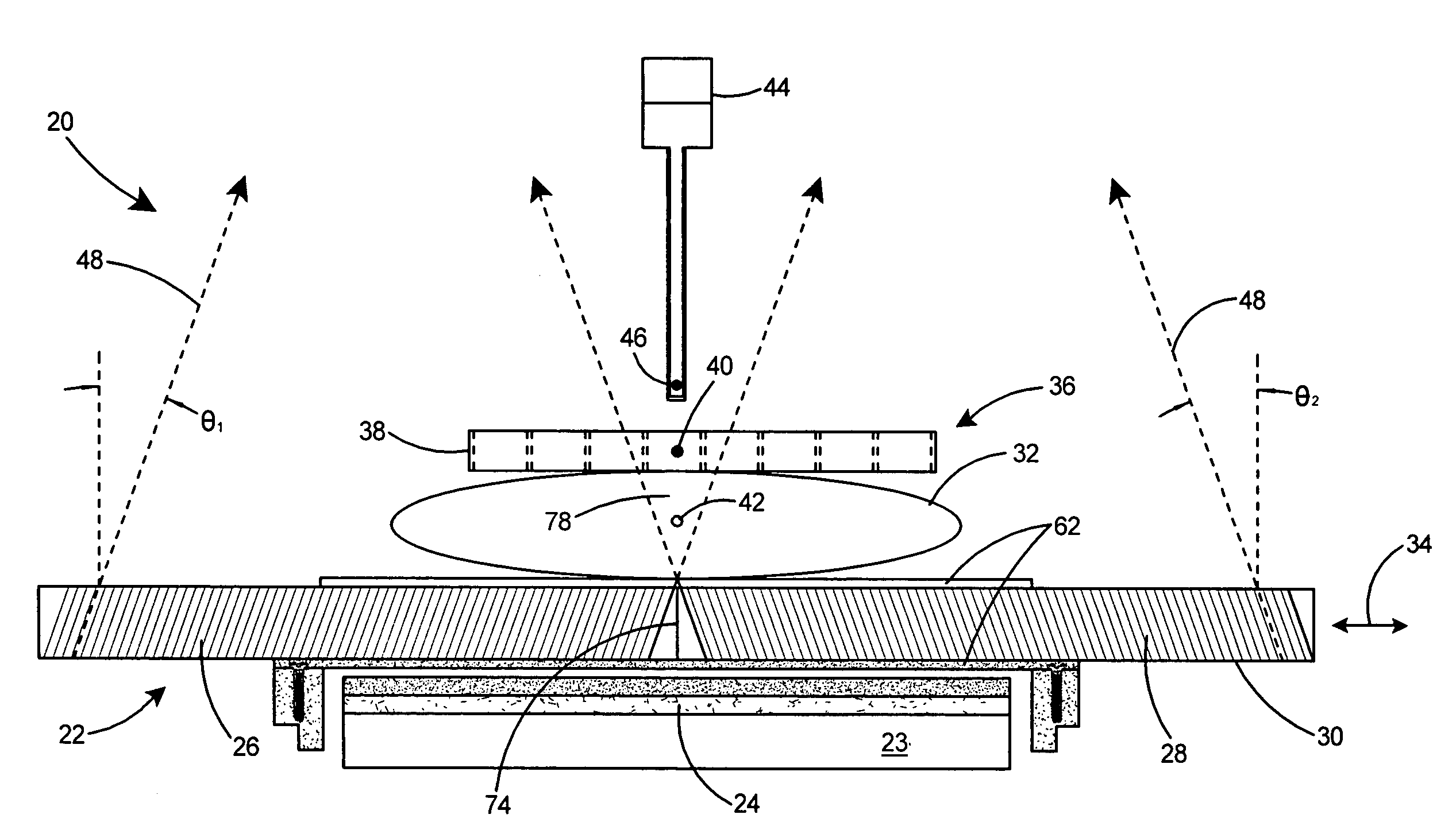
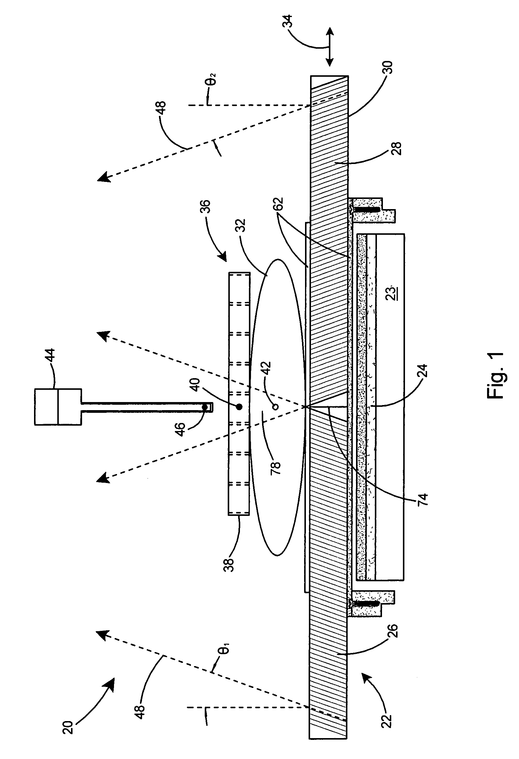
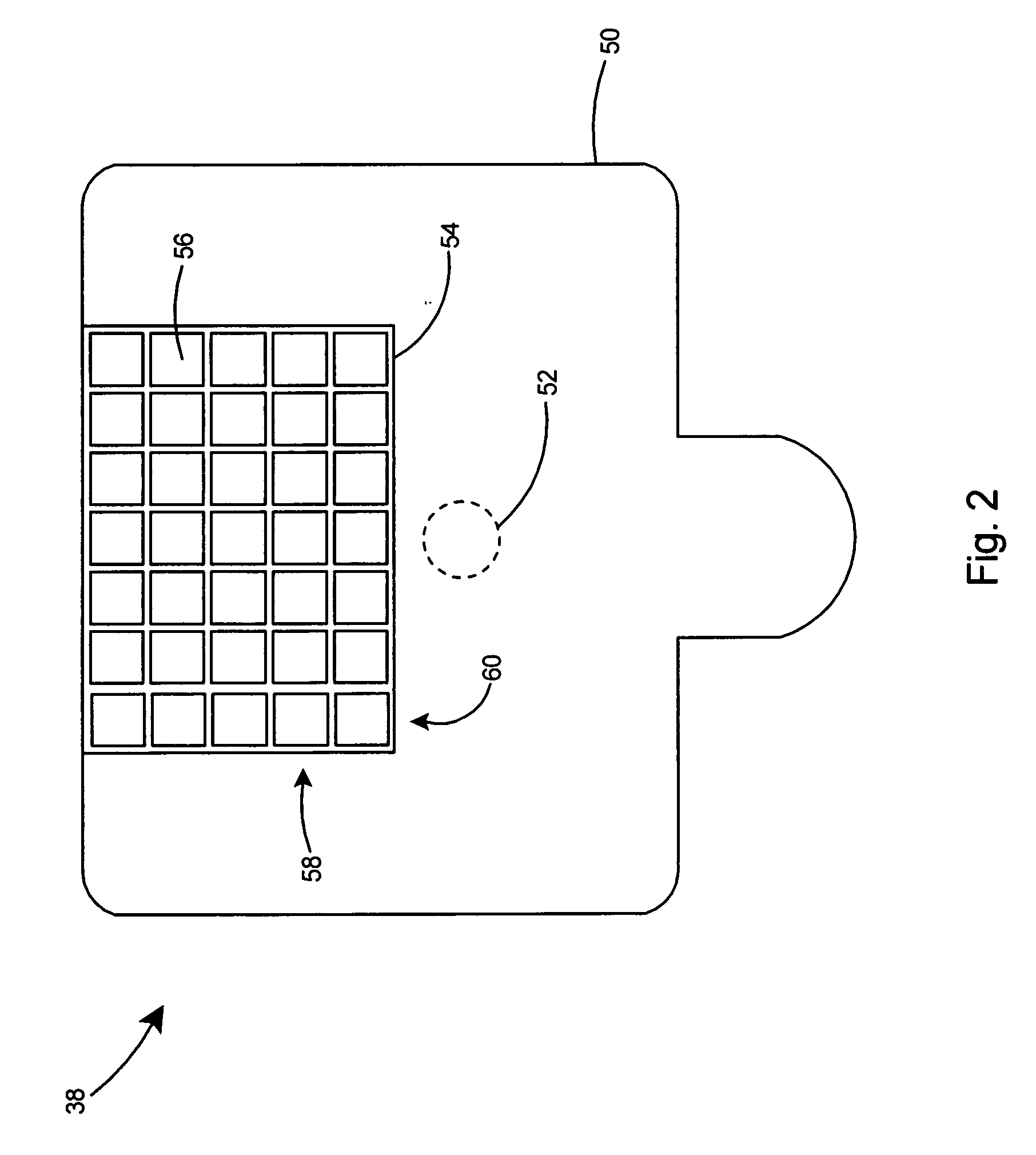
[0060]The present invention provides a gamma guided stereotactic localization system for accurately locating and guiding biopsy equipment to cancerous lesions. The gamma guided stereotactic localization system of the present invention is a functional or molecular breast imaging procedure that captures the metabolic activity of breast lesions through radiotracer uptake. A small amount of tracing agent is delivered to a patient, and in turn is absorbed by all cells in the body. The tracing agent emits invisible gamma rays, which are detected by a gamma camera and translated into a digital image of the breast. Due to the higher metabolic activity of cancerous cells, these cells absorb a greater amount of the tracing agent and are revealed as “hot spots.” This molecular breast imaging technique can help doctors more reliably differentiate cancerous from non-cancerous cells. While other adjunct modalities, such as MRI and ultrasound, image the physical structure of the breast, the gamma ...


PUM


Abstract

A stereotactic gamma-guided localization system for imaging a suspected cancer and guiding a physician in the removal of tissue samples for biopsy. The gamma-guided localization system includes a three step procedure including localization, correlation, and verification. The localization system includes a gamma camera with a set of slant-hole collimators for producing stereo images of a region of interest. A positioning system including a fiducial marker is placed adjacent to the object to be imaged and held rigidly in place to provide correlation of the location of the region of interest relative to the fiducial marker. A gamma emitting marker is then positioned at the calculated location of the region of interest and imaged to verify that that the calculated position corresponds to the actual location. The positioning system can then be used to accurately position and support any other hardware that needs to be positioned at the region of interest.

Description

FIELD OF THE INVENTION[0001]This invention relates to imaging of suspected cancer and specifically to a gamma imaging system used to guide a physician in the removal of tissue samples for biopsy.BACKGROUND OF THE INVENTION[0002]Mammographic imaging is well established as the primary screening modality for breast cancer. A suspicious finding on a mammographic examination may lead to imaging with another modality to further investigate the suspicious finding and ultimately to a biopsy being performed to confirm that cancer is or is not present. The other modalities may include a diagnostic mammogram, an ultrasound (US) examination, a magnetic resonance imaging (MRI) procedure, or a nuclear medicine procedure (known as scintimammography). Depending on the nature of the finding and the imaging system with which it was found, the surgeon or radiologist may be guided in the removal of tissue for pathological examination by one of these imaging systems. Breast biopsy systems have been prod...

Claims


Application Information

Patent Timeline
Patent Type & Authority Applications(United States)
IPC IPC(8): A61B19/00
CPCA61B19/5212A61B19/5244A61B6/4258A61B2019/542A61B2019/5227A61B2090/392A61B34/20A61B90/361A61B2090/371A61B5/00A61B5/055A61B6/00
Inventor KIEPER, DOUGLAS A.WELCH, BENJAMIN LAWRENCEFAIRCHILD, LEE HOLLAND
Owner DILON TECH
Why Eureka
Try Eureka