Hip Helical Implant

a technology of helical implants and hips, which is applied in the field of hip helical implants, can solve the problems of increasing the risk of bending or breaking the implanted hip nail, nail piercing the surface of the femoral neck or head, and inability to allow the fracture's bone fragments to slide towards each other along the hip nail, etc., and achieves minimal bone loss, easy insertion procedure, and minimal bone loss.

Active Publication Date: 2010-03-11
DEPUY SYNTHES PROD INC
View PDF10 Cites 14 Cited by
  • Summary
  • Abstract
  • Description
  • Claims
  • Application Information

AI Technical Summary

Benefits of technology

[0014]To obtain a simple insertion technique that permits inserting the partial helical implant without the loss of bone tissue, a step helix is developed. The step helix consists of at least two helically twisted blades (same number as the blades in the hip helical implant) attached to a base at its rear (proximal) end. During the insertion procedure, the step helix is solidly fixed to the hip helical implant, in such a way that when assembled one with the other, the combination constitutes a temporarily complete helical assembly. This permits that during insertion, the hip helical implant turns in a constant pace and rhythm, and the guidance by the slots at the intramedullary nail as it advances towards the femoral head until its final position diminishes the bone loss provoked during insertion. When insertion is completed, the step helix is removed, leaving only the hip helical implant inside the femoral neck, which, having a rear smooth shaft, is able to slide back inside the sliding sleeve, thus permitting fracture compression.
[0015]An insertion tool allows for an easier insertion procedure. The insertion tool consists of a cannulated shaft with a rotating handle that is able to turn around the shaft; an axial insert that goes inside the cannulation in the shaft; and a rear cap that engages the rear end of the axial insert. Before insertion, the step helix is solidly engaged with the hip helical implant, from behind, constituting the helical assembly. Thereafter, the insertion tool is attached to the rear end of the helical assembly. This permits a proper engagement of the insertion tool with the step helix and the hip helical implant enabling an appropriate insertion procedure.
[0016]During the insertion procedure, the rear end of the insertion tool is hammered, pushing the helical assembly forward through the oblique bore of the intramedullary nail (previously introduced in the medullary canal) towards the femoral head. The slots at the oblique bore guide the insertion of the helical assembly; this, together with the affixing of the step helix to the hip helical implant, allows the helical implant to turn in a constant pace and rhythm as it advances during the insertion procedure, causing the minimal bone loss.
[0017]After insertion is completed, the step helix and the insertion tool are easily removed., leaving the hip helical implant, and the sliding sleeve is introduced over the shaft of the hip helical implant and through the oblique bore of the intramedullary nail, permitting sliding back of the hip helical implant through the sleeve. This provides rotational stability due to the above described flats. Thereafter, a lateral set screw is inserted in the notch of the intramedullary nail affixing the sliding sleeve to the nail.

Problems solved by technology

This device, while permitting an adequate fixation and rotational control of the fracture, was unable to allow sliding of the fracture's bone fragments towards each other along the hip nail.
As a result, bone contact was insufficient to support a patient's weight, resulting in an increased risk of bending or breaking of the implanted hip nail.
This, together with the shape of the hip nail, determinate too much pressure over the femoral neck and head bone tissue, could lead the implant to cut through the cancellouse tissue of the femoral neck or head in a condition known as “cut out”, causing the nail to pierce the surface of the femoral neck or head, or at the least to lose proper alignment of the bone fracture.
Thus, when the healing bone is under the patients' weight, the implant may cut through the cancellous bone of the femoral head, causing the implant to rupture the femoral surface, or to no longer maintain a proper alignment of the fracture.
Another disadvantage of these types of implants is that they lack rotational control, permitting the rotation of the femoral head around the hip screw.
Although this may solve the cutting out problem and achieve adequate rotational stability, this system does not allow the sliding back of the implant through the vertical slot in the intramedullary nail, and therefore fails to permit the necessary bone fragment migration needed to provide fracture compression.
The shaft needed to permit sliding does not allow rotational control, which may result in the rotation of one bone fragment around another.
In addition, partial helical implants have an additional draw-back: the helical implant needs to be inserted in a guided way that permits the implant to rotate in a constant and predetermined rhythm, otherwise the implant would provoke femoral neck and head tissue loss while being inserted, and as a consequence the fracture fixation may become unstable, mainly in osteoporotic bones.
Such a design and use is very complex.

Method used


Image

  • Hip Helical Implant
  • Hip Helical Implant
  • Hip Helical Implant

Examples


Embodiment Construction

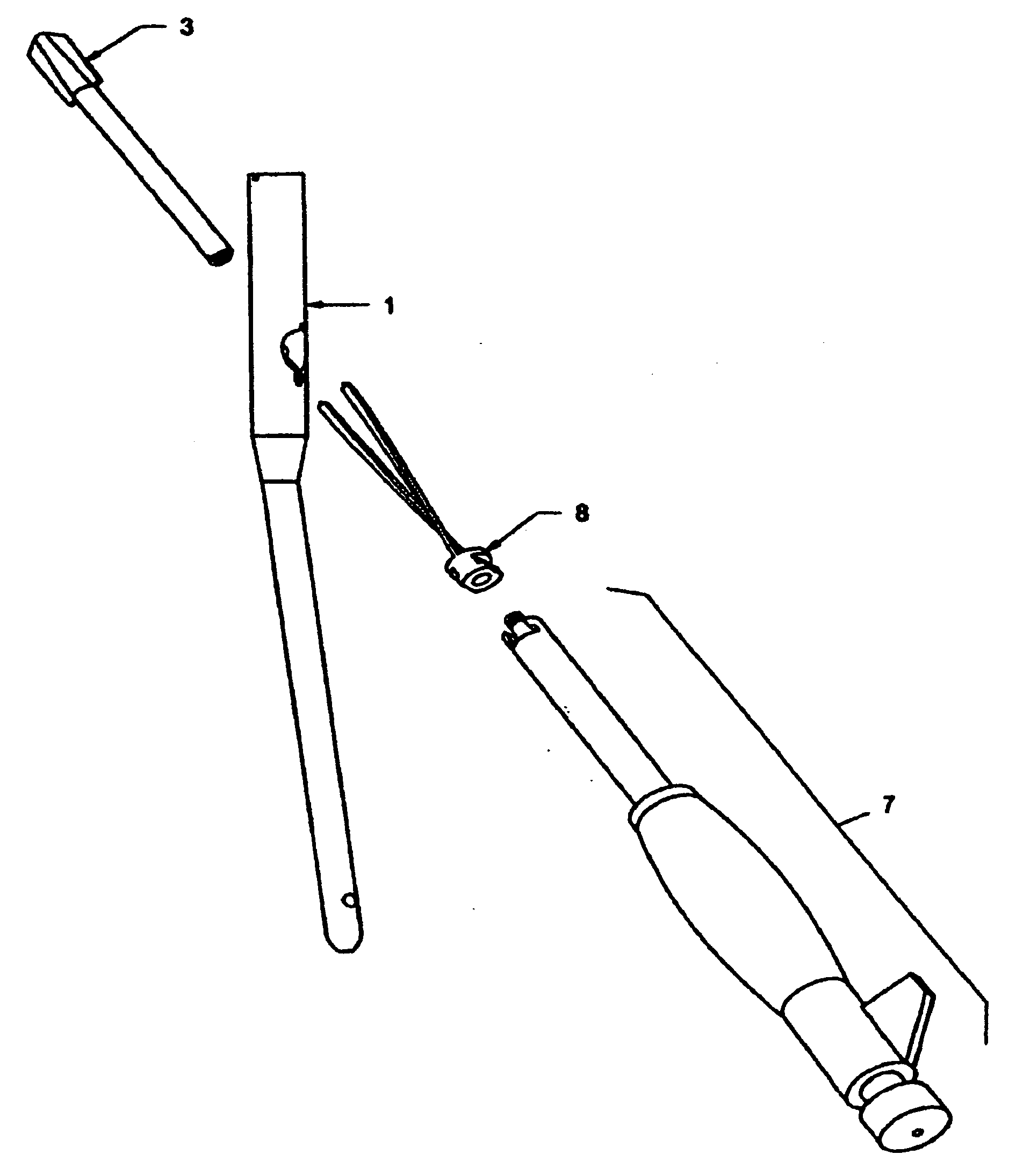
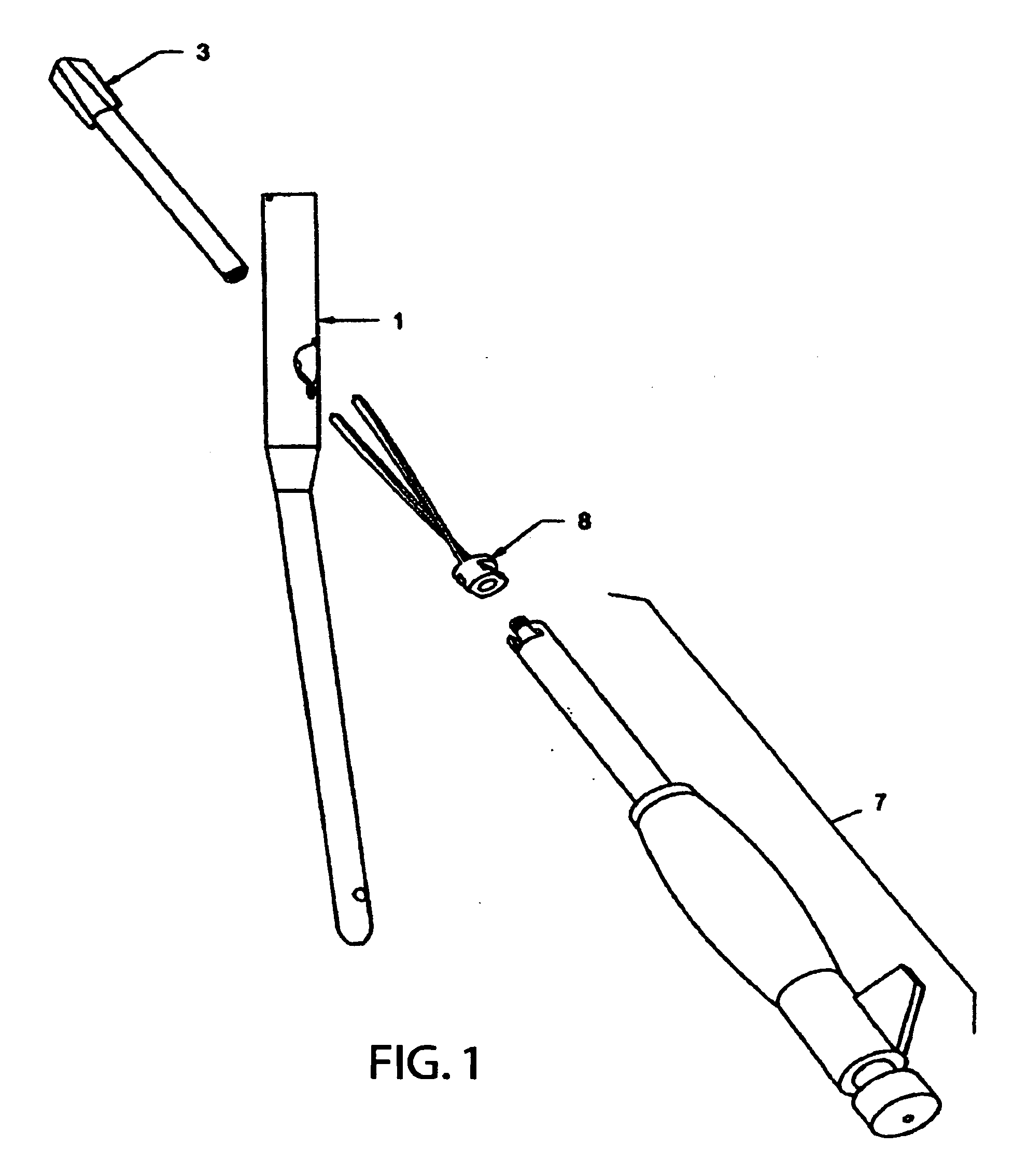
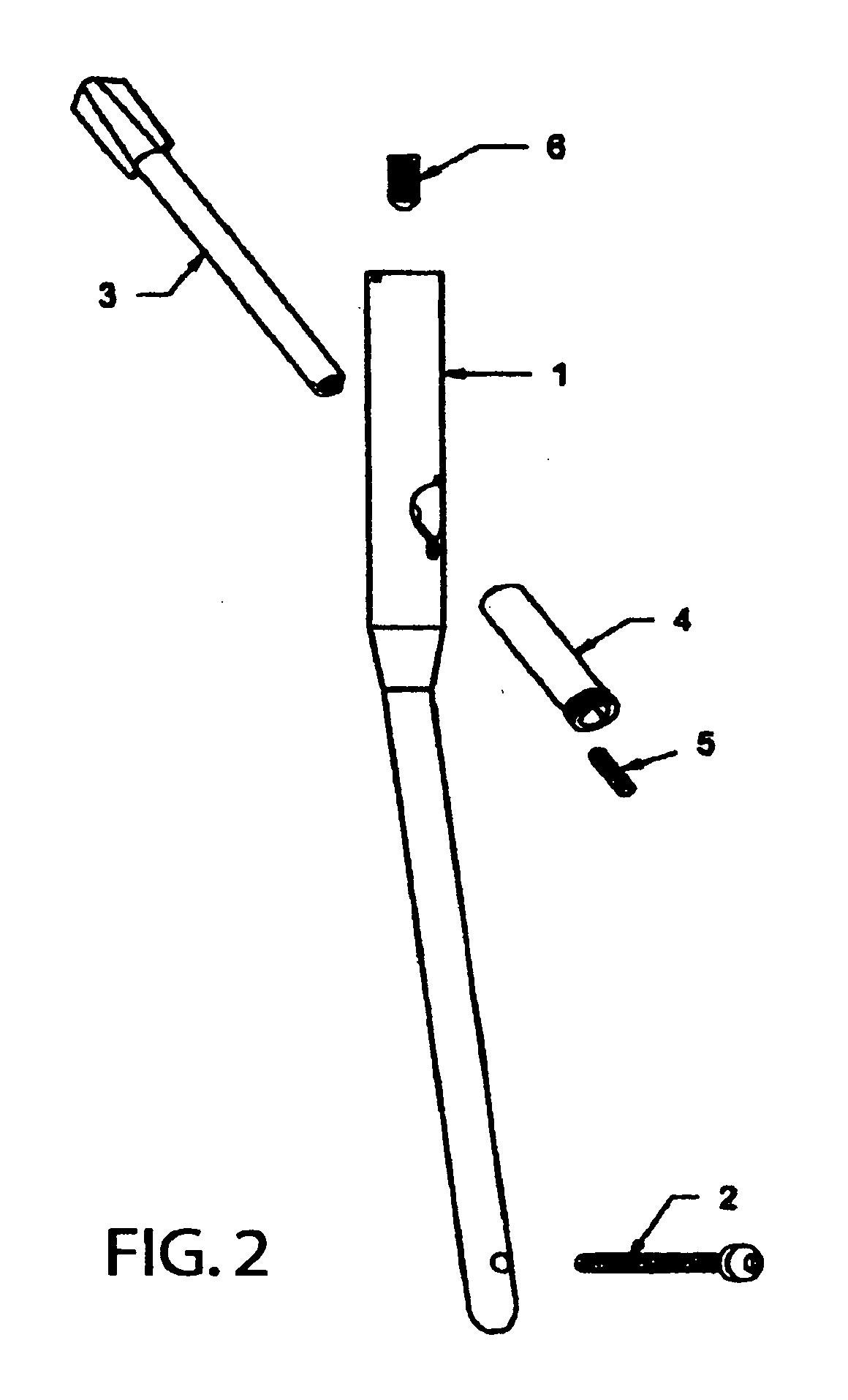
[0051]FIGS. 1 and 2 illustrate individual components of a preferred embodiment of the intramedullary ostesynthetic device, which includes an intramedullary nail 1 with optional distal locking screws 2, a hip helical implant 3, a sliding sleeve 4, a lateral set screw 5, and an optional coaxial set screw 6; as well as an insertion tool 7 and a step helix 8. The insertion tool 7 and step helix 8 may be used during the insertion procedure.

[0052]FIGS. 3 to 3B illustrate a preferred embodiment of the hip helical implant 3, which consists of a front helical portion 11, and a rear smooth shaft 9. The frontal helical portion 11 is provided of at least two helically twisted blades 10, which are attached to the axis of the front helical portion 11 that has a truncated conical shape. Additional blades are contemplated. Each blade 10 is provided with a hole 12 at its rear end, designed to receive the pegs 30 located at the frontal ends of the step blades 28 of the step helix 8 (FIG. 7), in order...


PUM


Abstract

An intramedullary osteosynthetic device includes an intramedullary nail, a hip helical implant, a sliding sleeve, a lateral set screw, and at least one distal locking screw. The intramedullary nail is cannulated. The cannulation is at least partially threaded to receive a coaxial set screw. The intramedullary nail includes at least one oblique slotted bore in communication with the cannulation, where the at least oblique slotted bore receives a sliding sleeve, and further includes a threaded notch compatible with the lateral set screw. The hip helical implant includes a frontal helical portion having a truncated conical shape, at least two helical twisted blades attached to the frontal helical portion, and a rear smooth shaft, the shaft having an external flat surface. Each blade of the hip helical implant has a hole for receiving a peg from a step helix fixating the hip helical implant to the step helix. The hip helical implant is cannulated to receive a Kirschner wire during insertion of the implant into a femoral head. The sliding sleeve includes a tube having an internal flat corresponding to the external flat of the hip helical implant and has external threads at the rear of the sleeve engaging the lateral set screw. The coaxial set screw fixes the sliding sleeve to the intramedullary nail by tightening the sliding sleeve inside the oblique bore.

Description

FIELD OF THE INVENTION[0001]The present invention relates to a device for use in osteosynthesis to treat femoral fractures, in particular fractures of the neck and the intertrochanteric region of the femur.BACKGROUND OF THE INVENTION[0002]Many implants have been developed to treat intertrochanteric femoral fractures which are basically based on a hip nail or screw that is inserted from the side of the femur through the neck and into the femoral head, and afterwards it is fixed either to an intramedullary nail positioned inside the femoral shaft, or to a side plate positioned in the outside of the femoral shaft.[0003]In 1969 Zickel developed an intramedullary rod and cross nail assembly. U.S. Pat. No. 3,33,220 discloses a hip nail fixed to an intramedullary nail inside the femoral shaft. This device, while permitting an adequate fixation and rotational control of the fracture, was unable to allow sliding of the fracture's bone fragments towards each other along the hip nail. As a res...

Claims


Application Information

Patent Timeline
Patent Type & Authority Applications(United States)
IPC IPC(8): A61B17/56A61B17/58
CPCA61B17/744A61B17/862A61B2017/8655A61B17/8883A61B17/8891A61B17/8685A61B17/72A61B17/74
Inventor DELL'OCA, ALBERTO A. FERNANDEZ
Owner DEPUY SYNTHES PROD INC
Why Eureka
Try Eureka