

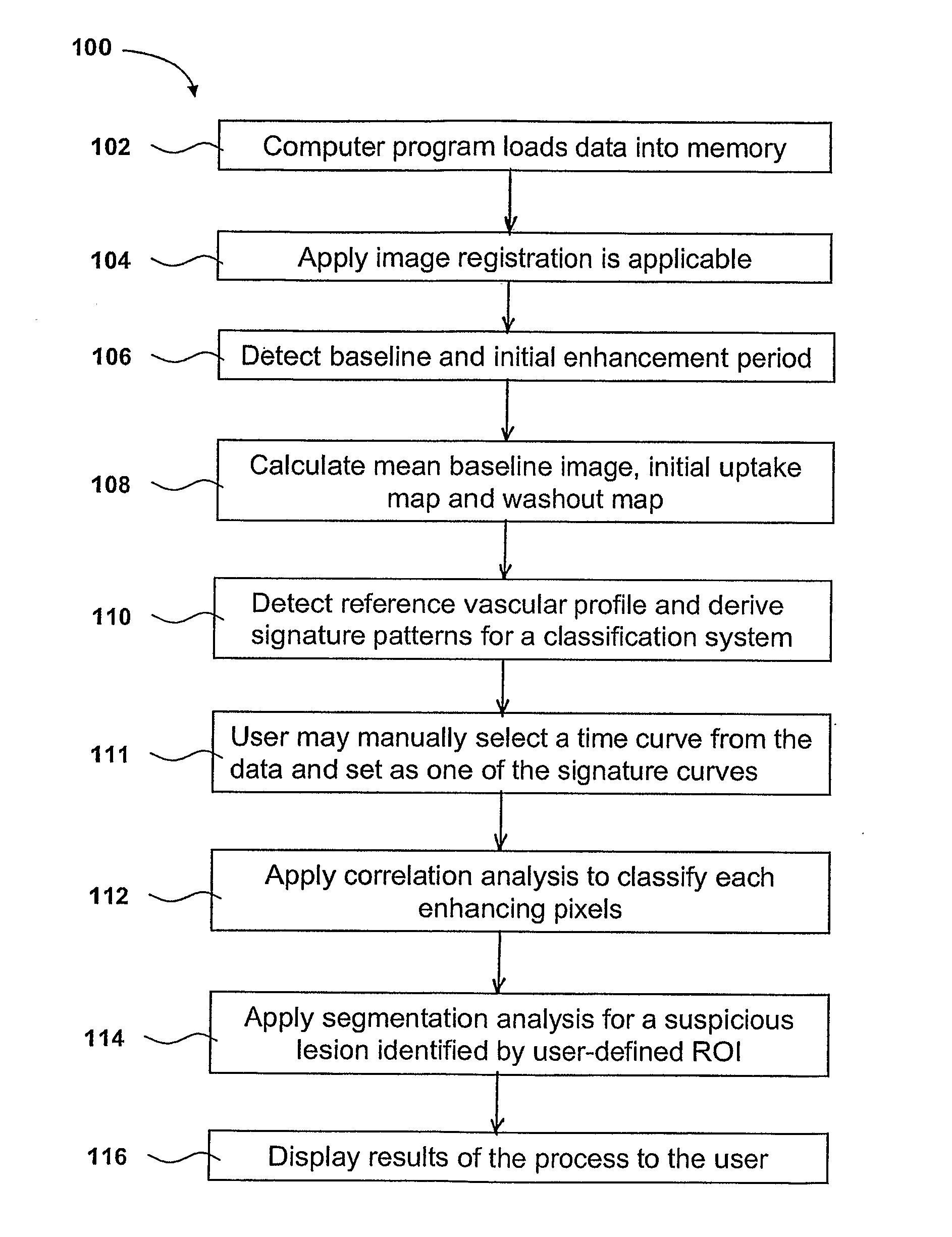
Identification and analysis of lesions in medical imaging
a technology for diagnosing and analyzing lesions, applied in image data processing, diagnostics, applications, etc., can solve the problems of not providing statistical significance information associated, based on calculated descriptive parameters, and time-consuming for medical personnel to review all images
- Summary
- Abstract
- Description
- Claims
- Application Information
AI Technical Summary
Benefits of technology
Problems solved by technology
Method used
Image

Examples
Embodiment Construction
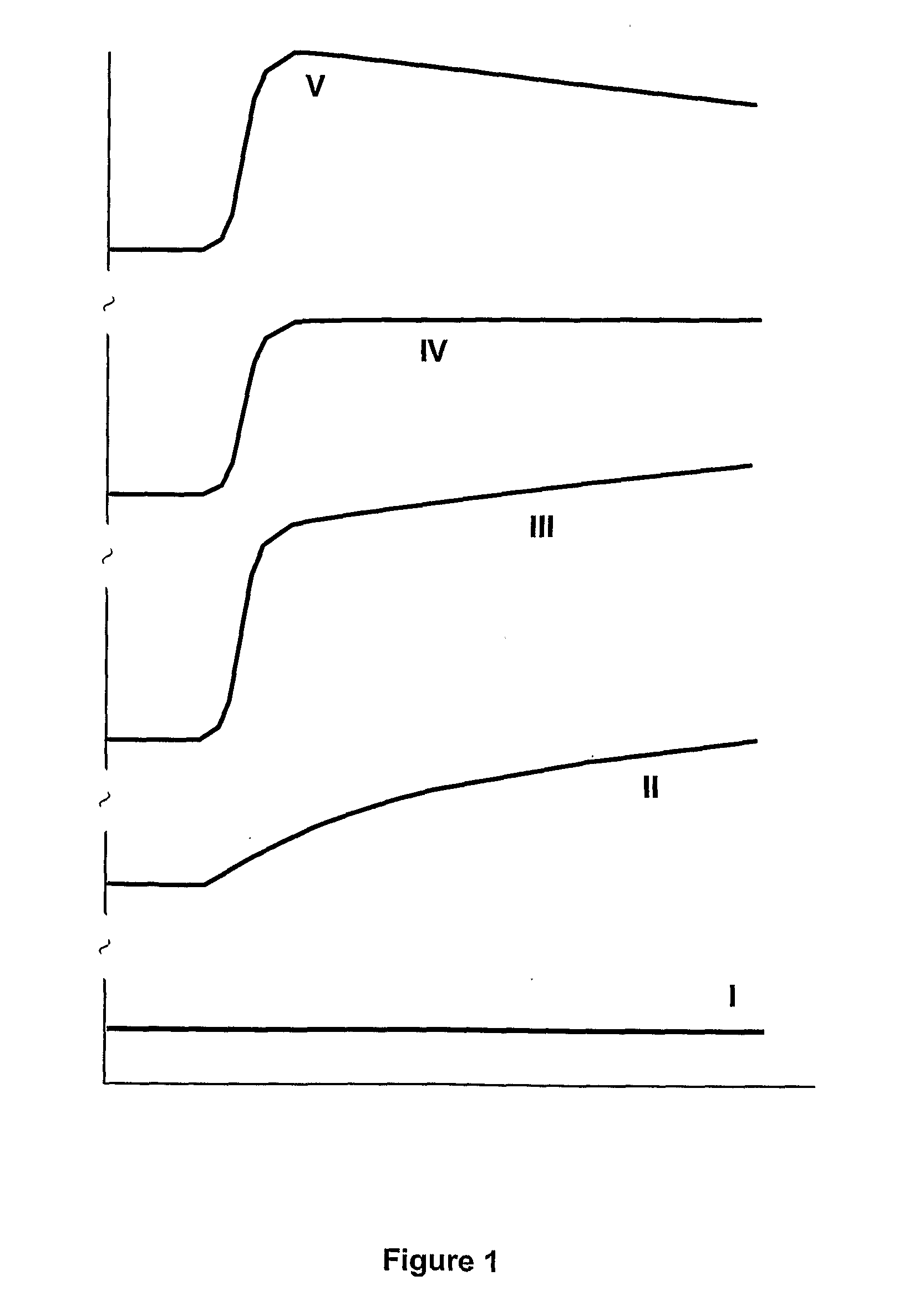
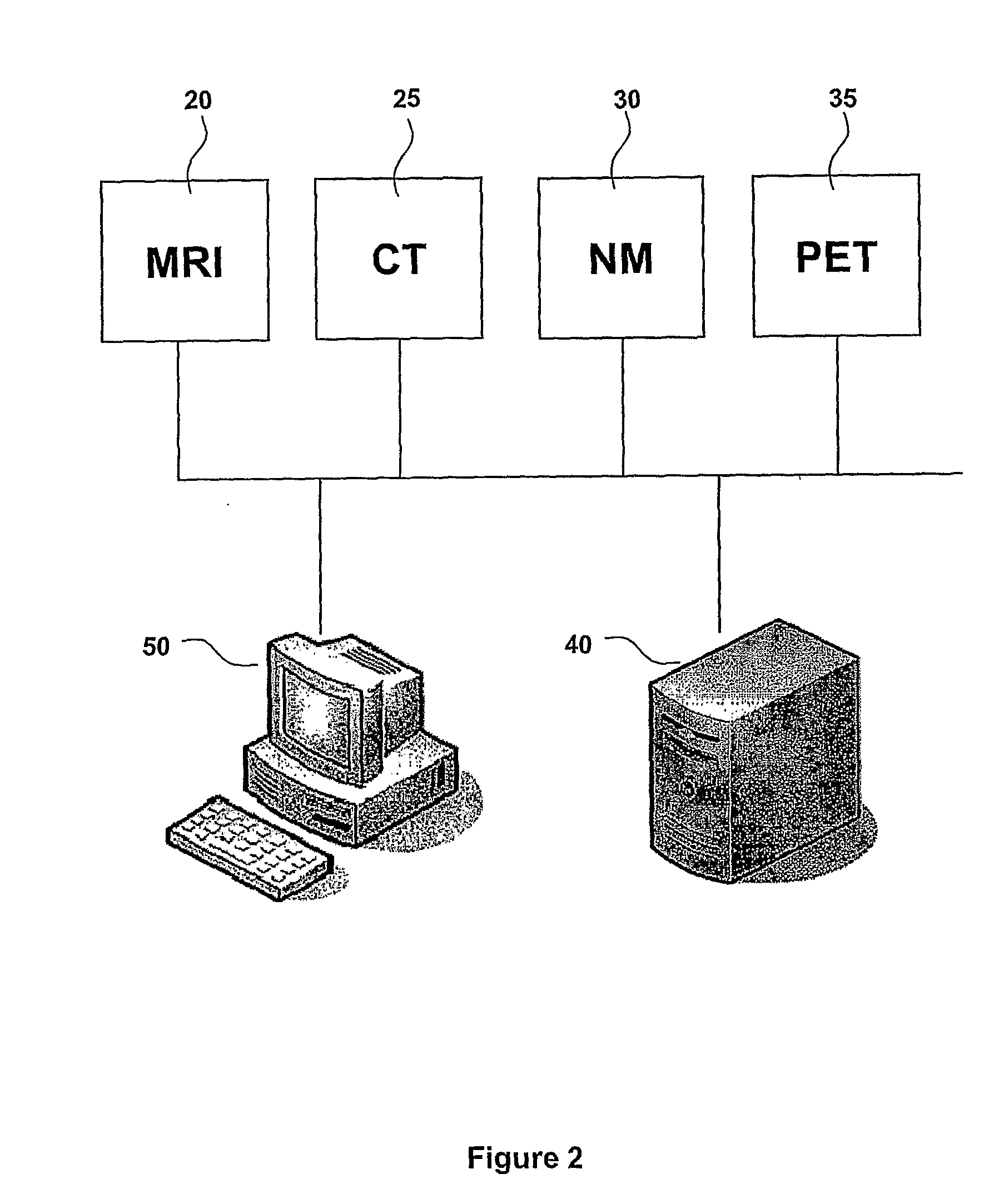
[0056]The present invention is particularly applicable to dynamic MRI, CT, NM and PET imaging systems, for example dynamic MRI of the breast. Raw data and / or images collected by a scan, such as from a MRI scanner 20, CT scanner 25, NM scanner 30 or PET scanner 35 are forwarded to a data storage system 40 in the form of a Picture Archiving Communications System (PACS) in FIG. 2. A computer program operating on a processor 50, in the form of a computer, is used to retrieve or receive the various images or raw data from any one of the scanners 20, 25, 30 or 35 or from the data storage system 40. The program then processes those images to provide an improved data set for medical personnel to use, particularly in relation to evaluate both morphologic and kinetic information for diagnosis of disease and optimal discrimination between benign and malignant disease. The computer program need not reside on computer 50, but may reside in a console computer linked to any one of the scanners 20,...
PUM
 Abstract
Description
Claims
Application Information