System for multi-dimensional anatomical functional imaging

a multi-dimensional anatomical and functional imaging technology, applied in the field of multi-dimensional anatomical functional imaging, can solve the problems of unnecessarily exposing patient tissue and organs to additional radiation, time-consuming, subjective clinical evaluation of frames,

Inactive Publication Date: 2010-04-01
SIEMENS HEALTHCARE GMBH
View PDF22 Cites 94 Cited by
  • Summary
  • Abstract
  • Description
  • Claims
  • Application Information

AI Technical Summary

Problems solved by technology

However, the clinical evaluation of the frames may be subjective, time-consuming and requires extensive expertise and clinical experience for accurate interpretation and proper cardiac rhythm management.
Additionally, continuous image scanning and data acquisition may unnecessarily expose patient tissue and organs to

Method used


Image

  • System for multi-dimensional anatomical functional imaging
  • System for multi-dimensional anatomical functional imaging
  • System for multi-dimensional anatomical functional imaging

Examples


Embodiment Construction

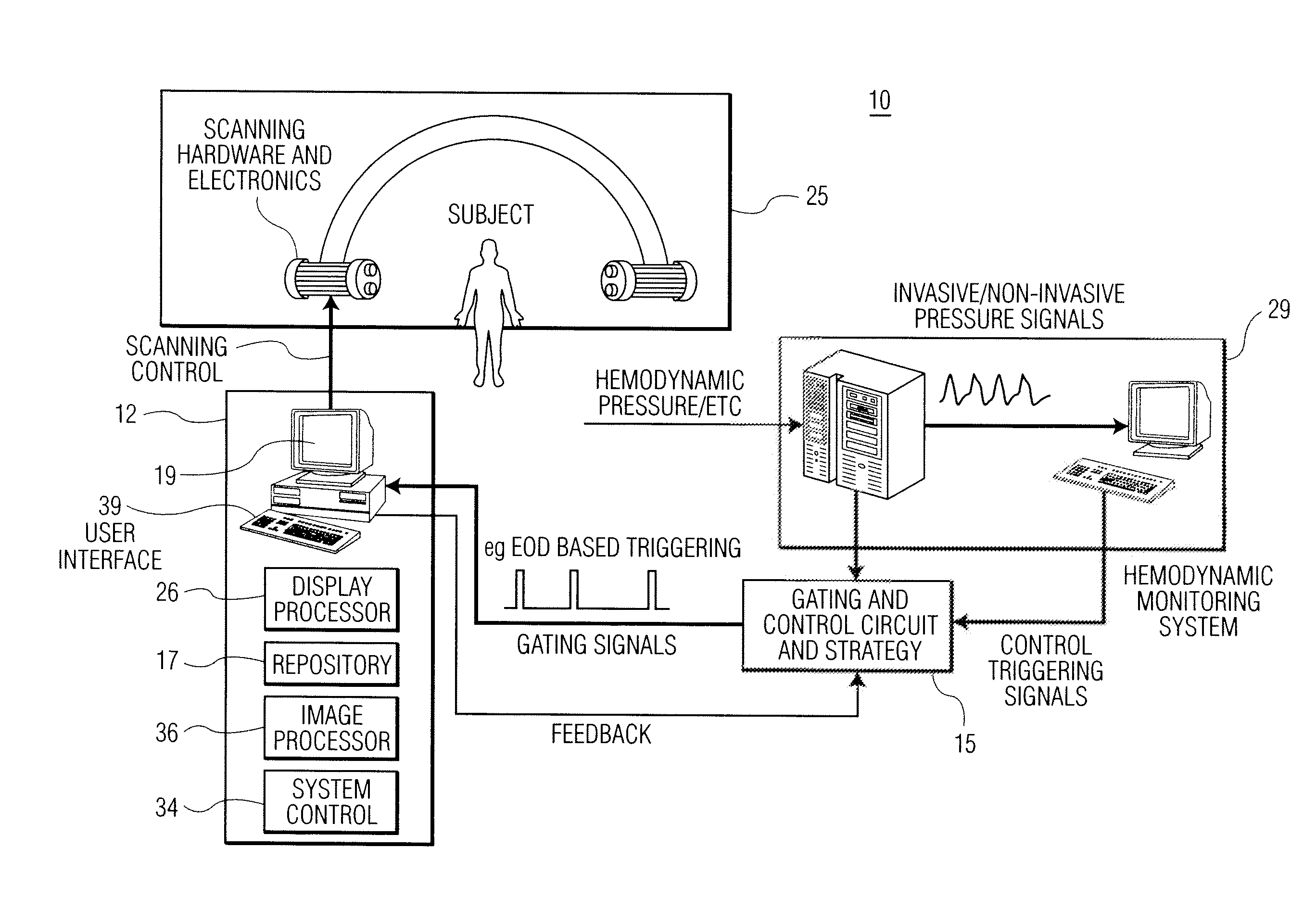
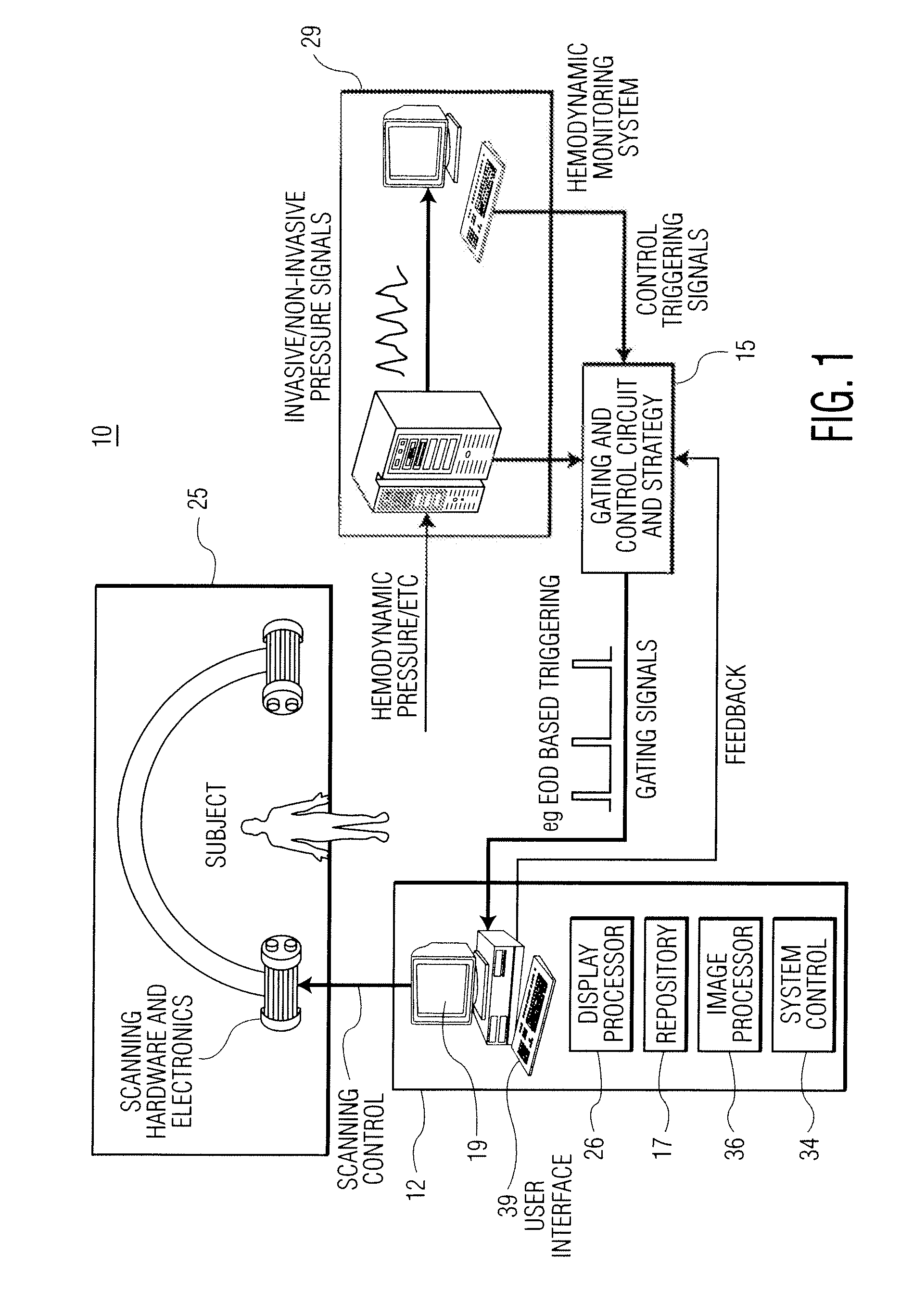
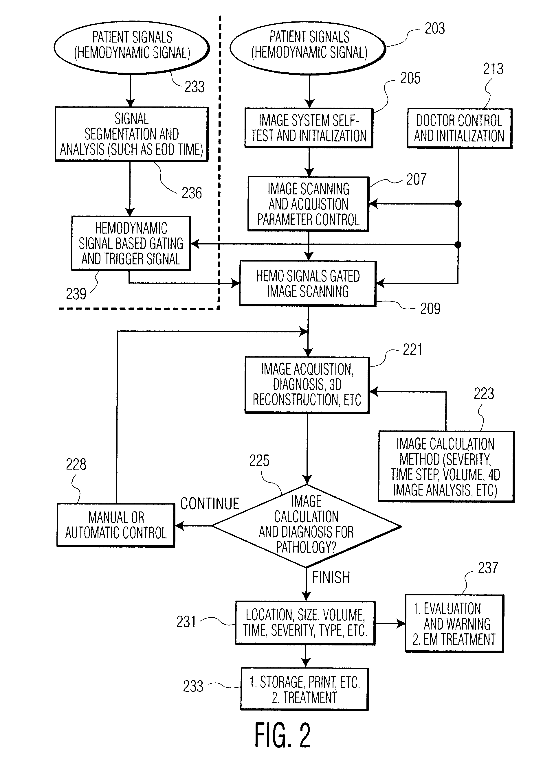
[0011]A cardiac functional analysis system improves image quality and scanning efficiency in X-ray image acquisition using a hemodynamic signal output. The acquisition and imaging unit acquires rotational images in response to a trigger signal derived from a hemodynamic signal (such as invasive blood pressure signal, blood volume calculation index) at predefined cardiac phases over multiple cardiac cycles. Data comprising 2D images is processed to provide multiple 3D volumes, with individual volumes constructed from frames acquired at the same cardiac phase over multiple cardiac cycles. The system provides a dynamic 4D cardiac dataset series for cardiac function assessment. The system provides more efficient safer use of an imaging system (such as an X-ray image system) with less power usage and radioactivity exposure.

[0012]The system employs hemodynamic signal based image gating and acquisition using cardiac hemodynamic signals to synchronize image scanning for cardiac tissue and f...


PUM


Abstract

A cardiac functional analysis system reconstructs a 3D anatomical image volume using image frames acquired at predetermined cardiac phases over multiple cardiac cycles in response to a trigger derived from hemodynamic signals. A medical imaging system generates 3D anatomical imaging volume datasets from acquired 2D anatomical images. The system includes an image acquisition device for acquiring 2D anatomical images of a portion of patient anatomy in selectable angularly variable imaging planes in response to a synchronization signal derived from a patient blood flow related parameter. A synchronization processor provides the synchronization signal derived from the patient blood flow related parameter. An image processor processes 2D images acquired by the image acquisition device of the portion of patient anatomy in multiple different imaging planes having relative angular separation, to provide a 3D image reconstruction of the portion of patient anatomy.

Description

[0001]This is a non-provisional application of provisional application Ser. No. 61 / 101,373 filed Sep. 30, 2008, by H. Zhang et al.FIELD OF INVENTION[0002]This invention concerns a system for generating 3D anatomical images from acquired 2D anatomical images in response to a synchronization signal derived from a patient blood flow related parameter.BACKGROUND OF INVENTION[0003]The evaluation of heart health status normally involves analyzing several functional characteristics of a left ventricle. These characteristics include ejection fraction, cardiac output, stroke volume, wall motion and wall thickness. The evaluation of heart health status may also include analysis of a right ventricle and atrium. Cardiac function analysis also usually involves image scanning a patient heart chamber of interest using an X-ray imaging device over multiple heart cycles. A contrast agent (dye) is injected into a patient to improve visualization of a chamber and the flow of the blood pool in and out ...

Claims


Application Information

Patent Timeline
IPC IPC(8): A61B5/05G06K9/00
CPCA61B6/503A61B6/5217A61B6/5288G06T2211/412A61B8/5284G06T11/006A61B6/541G16H50/30
Inventor ZHANG, HONGXUANCHEN, JINGHUA
Owner SIEMENS HEALTHCARE GMBH
Why Eureka
Try Eureka