Nanoparticles for Delivery of Therapeutic Agents Using Ultrasound and Associated Methods

a technology of nanoparticles and therapeutic agents, applied in the field of lipid-based nanoparticles or liposomes, can solve the problems of limited amount of active agents that can be delivered, inability to visualize the targeted region while at the same time controlling the release of payloads, etc., to reduce the time required for treatment, reduce the circulating level and amount of active agents, and reduce the effect of pharmaceutical production costs

Inactive Publication Date: 2010-05-27
WATKIN KENNETH L
View PDF10 Cites 1 Cited by
  • Summary
  • Abstract
  • Description
  • Claims
  • Application Information

AI Technical Summary

Benefits of technology

[0010]Another object of the present invention is to reduce the circulating levels and amounts of active agents that are required for treatment by providing particles capable of encapsulating greater amounts of the active agents.
[0011]Another object of the present invention is to reduce the time required for treatment by providing a method for releasing encapsulated agents using ultrasound.
[0012]A further object of the present invention is to lower pharmaceutical production costs by providing particles capable of encapsulating greater amounts of the active agents.

Problems solved by technology

However, one of the drawbacks to using pH and / or temperature as the release mechanism is the inability to visualize the targeted region while at the same time controlling the release of the payload.
Another problem with current microspheres, liposomes, and nanoparticles is that they are formed using perfluorocarbon gas; the active agents are bound on the surface of the particle or within the layers forming the membrane or shell of the particle.
The amount of active agent that can be delivered is limited to the amount that is adhered to the surface of the particle or bound into the shell.

Method used


Image

  • Nanoparticles for Delivery of Therapeutic Agents Using Ultrasound and Associated Methods
  • Nanoparticles for Delivery of Therapeutic Agents Using Ultrasound and Associated Methods
  • Nanoparticles for Delivery of Therapeutic Agents Using Ultrasound and Associated Methods

Examples


example 1

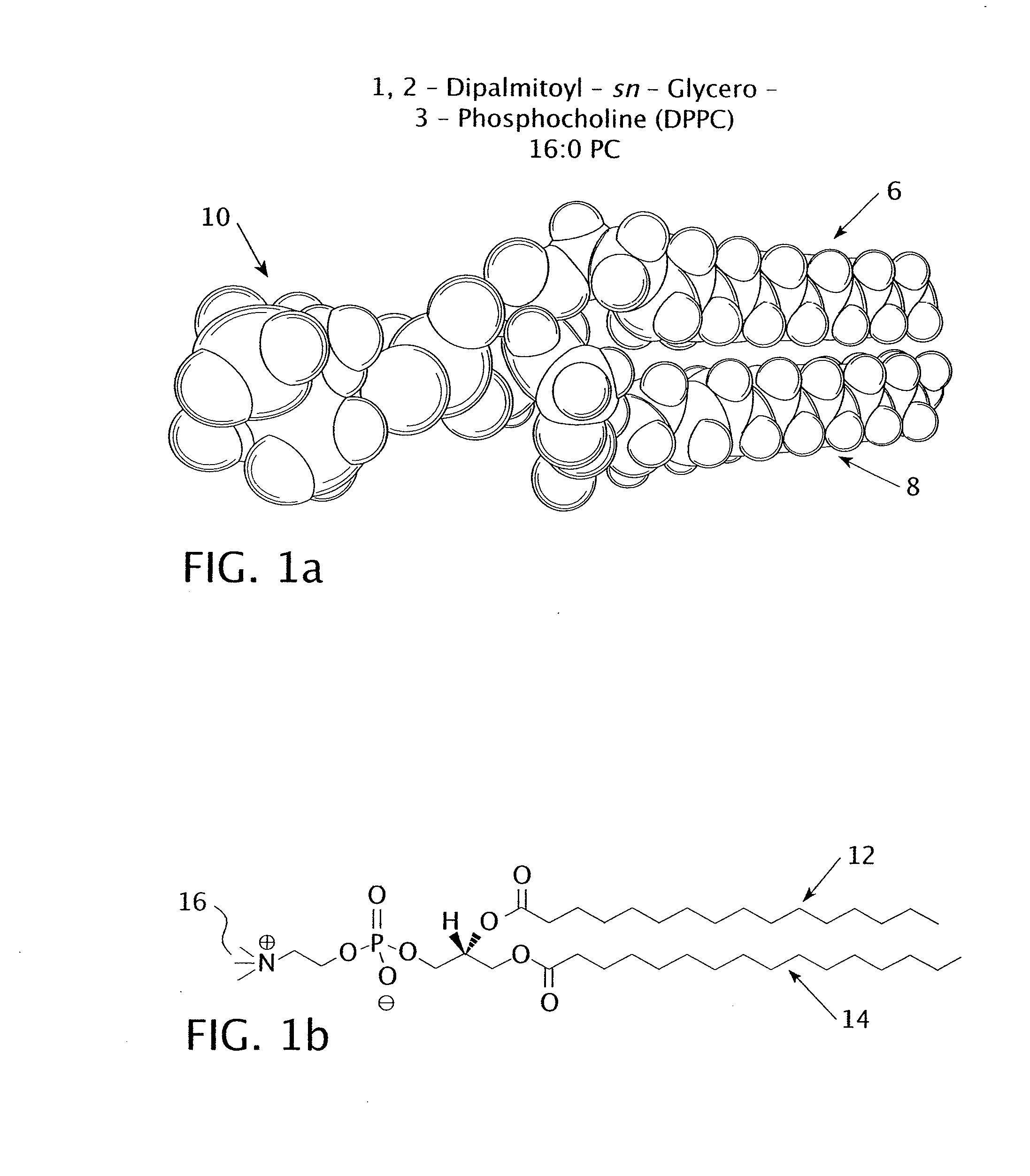
[0037]The experiment tested the percent release of encapsulated carboxyfluorescein from three different types of nanoparticles. The first type of nanoparticle included a DPPC bilayer with no lysolipid; the DPPC had a chain length of 16. The second type of nanoparticle included a DPPC and MMPC bilayer with a molar ratio of 95:5 DPPC to MMPC. The DPPC had a chain length of 16 and the MMPC had a chain length of 14, giving the bilayer a chain length difference of 2. The third type of nanoparticle included a DPPC and MPPC bilayer with a molar ratio of 95:5 DPPC to MPPC. The DPPC and the MPPC had identical chain lengths of 16 each.

[0038]The amount of liquid carboxyfluorescein released from the particles was measured using a fluorometer. The nanoparticles were injected in a thin (2 mm) closed transparent membrane containing 2.5 ml of nanoparticles mixed with a buffered solution. Control samples were reserved prior to ultrasonic visualization and insonation for measurement purposes. The clo...

example 2

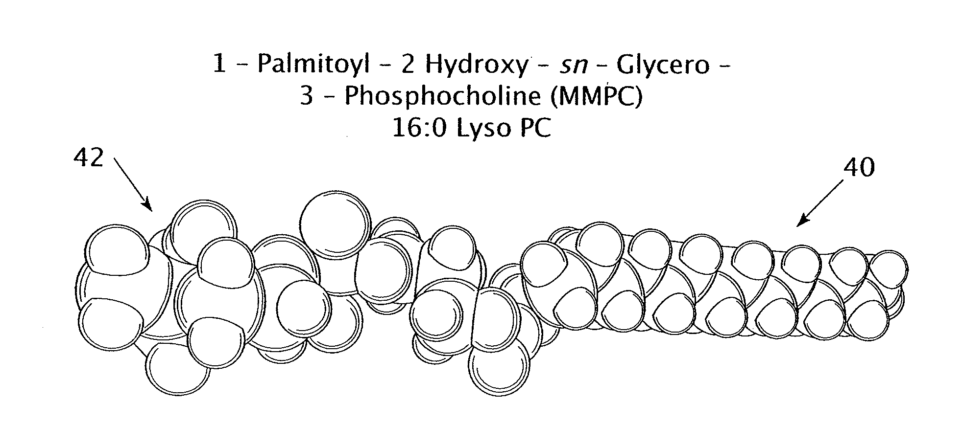
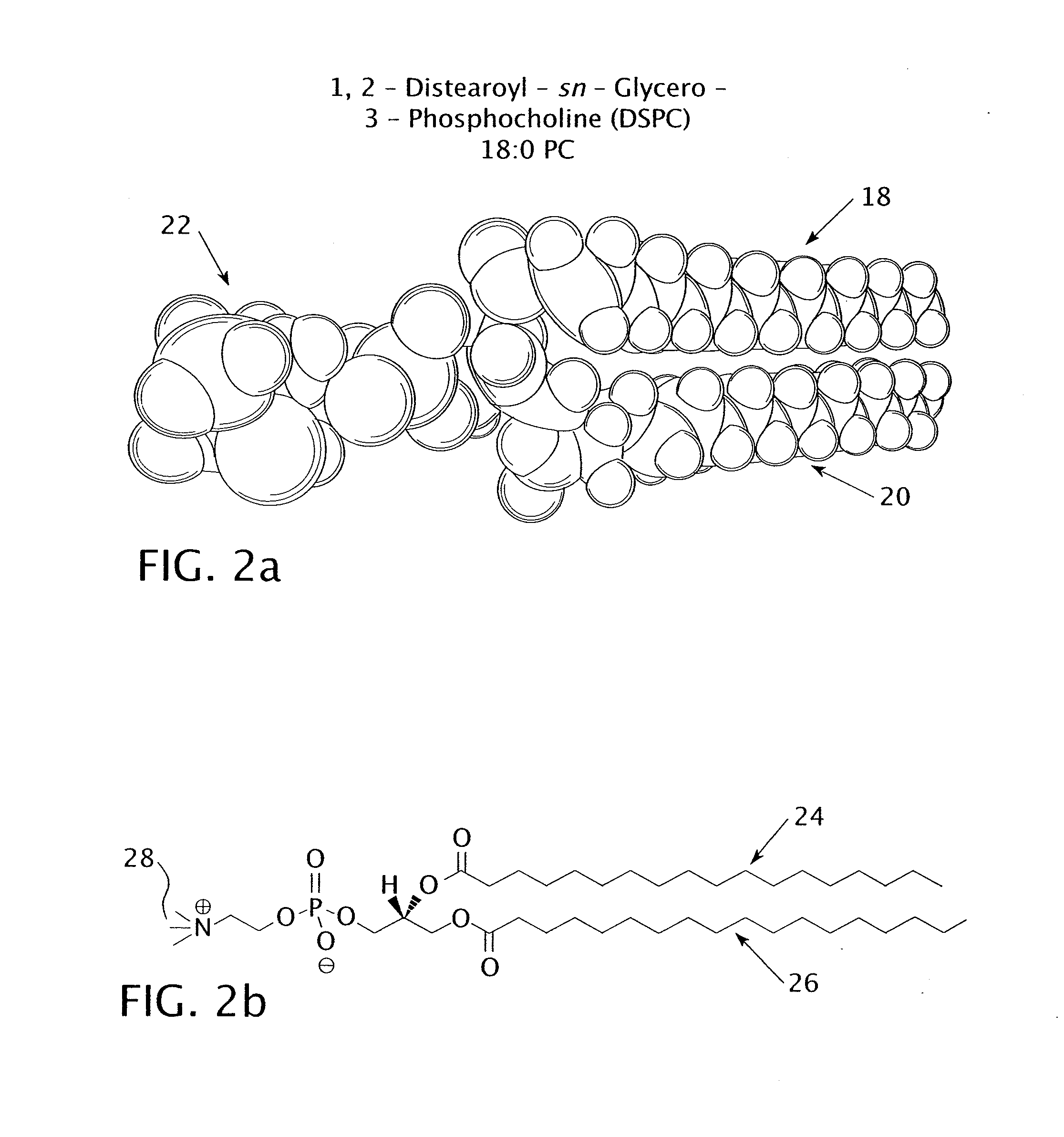
[0040]The experiment tested the percent release of encapsulated carboxyfluorescein from three different types of nanoparticles. The first type of nanoparticle included a DSPC bilayer with no lysolipid; the DSPC had a chain length of 18. The second type of nanoparticle included a DSPC and MMPC bilayer with a molar ratio of 95:5 DPPC to MMPC. The DSPC had a chain length of 18 and the MMPC had a chain length of 14, giving the bilayer a chain length difference of 4. The third type of nanoparticle included a DPPC (16:0) and MPPC (16:0) bilayer with a molar ratio of 95:5 DPPC to MPPC. The DPPC and the MPPC had identical chain lengths of 16 carbon atoms each.

[0041]The amount of liquid carboxyfluorescein released from the particles was measured using a fluorometer. The nanoparticles were injected in a thin (2 mm) closed transparent membrane containing 2.5 ml of nanoparticles mixed with a buffered solution. Control samples were reserved prior to ultrasonic visualization and insonation for me...

example 3

[0043]The following protocol was used to prepare a particle encapsulating carboxyfluorescein as the active agent:

[0044]1. Dissolve a primary phospholipid and a lysolipid in a 10 mg / mL solution of chloroform, which assists in preventing the formation of lipid spheres.

[0045]2. Calculate the required volume of liquid required to form a lipid bilayer based on molar percentages. For example:

[0046]Total concentration of the (DPPC:MMPC) lipid in a 95:5:10 umoL

95 / 100*10=9.5 umoL

5 / 100*10=0.5 umoL

[0047]DPPC volume is: (9.5E-6 moL)*(734.05 g / moL)*(10 L / g)=697 uL

[0048]MMPC volume is: (0.5E-6 moL)*(467.58 g / moL)*(10 L / g)=23.3 uL

[0049]3. Pipette the calculated volumes of the primary phospholipid and lysolipid in a container, e.g., a round bottom flask.

[0050]4. Blow a thin stream of N2 gas into the flask for approximately 2 seconds.

[0051]5. Seal the flask with parafilm and store in a freezer at approximately −20 degrees Celsius.

[0052]6. Dry the lipid solution by blowing a thin stream of N2 gas int...


PUM


Abstract

The present invention relates to lipid based nanoparticles or liposomes that are sensitive to ultrasonic energy, compositions containing these particles, methods for delivering one or more active agents using the particles, and methods for preparing the particles. The nanoparticles and liposomes encapsulate active agents such as chemotoxins, genes, virus vectors, proteins, peptides, antisense oligonucleotides, carbohydrates, and stem cells. The particles contain an aqueous core, at least one active agent located within the aqueous core, and a lipid bilayer or membrane that encapsulates the active agent within the aqueous core. The lipid bilayer may comprise a primary phospholipid and a lysolipid that preferably have different acyl chain lengths, making the lipid bilayer sensitive to ultrasound. Ultrasound may be used to track the particles as they move throughout the body. When the ultrasonic energy reaches a certain pressure, the lipid bilayer will break apart, releasing the active agent.

Description

FIELD OF THE INVENTION[0001]The present invention relates to lipid based nanoparticles or liposomes that are sensitive to ultrasonic energy, and more particularly relates to the use of these nanoparticles or liposomes to carry and release active agents such as chemotoxins, genes, virus vectors, proteins, peptides, antisense oligonucleotides, carbohydrates, and stem cells.BACKGROUND INFORMATION[0002]Nanoparticles and liposomes serve as ideal carriers for the delivery of payloads such as chemotoxins, genes, virus vectors, proteins, peptides, antisense oligonucleotides, carbohydrates, stem cells, and / or other agents for the treatment of disease. In general, a nanoparticle or liposome is a spherical vesicle with a membrane composed of a lipid bilayer that encapsulates an active agent, e.g., a therapeutic agent such as a drug or genetic material. When the lipid bilayer dissolves or breaks apart, the active agent is released into the body. Nanoparticles have a width of less than about 500...

Claims


Application Information

Patent Timeline
Patent Type & Authority Applications(United States)
IPC IPC(8): B01J13/04
CPCA61K9/127A61K9/0009
Inventor WATKIN, KENNETH L.
Owner WATKIN KENNETH L
Why Eureka
Try Eureka