Extending the resolution of MRI data by combining subsets from plural image acquisitions

a technology of mri data and subsets, applied in the field of nuclear magnetic resonance imaging, can solve the problem that the complete volumetric image would present a minimum time limit between stop frames, and achieve the effect of improving the extent, favorable short sampling time, and high light and contras

Inactive Publication Date: 2010-06-10
AURORA HEALTHCARE US CORP
View PDF24 Cites 15 Cited by
  • Summary
  • Abstract
  • Description
  • Claims
  • Application Information

AI Technical Summary

Benefits of technology

[0025]An important application of the disclosed technique is the diagnosis and treatment of breast cancer. By distinguishing tissue types based on their component elements or molecules, for example distinguishing concentrations of fat from concentrations of water, distinctions can be drawn to enable visualization of internal breast tissue structures, such as ducts and vasculature. Rendering fatty tissues transparent in a volume projection and enhancing water concentrations tends to highlight and impart contrast to the appearance of lesions in the images, helping a practitioner distinguish cysts from tumors, and so forth. Perfusing tissues with contrast agents improves the extent to which pertinent tissue types and tissue structures can be distinguished. Contrast agents assume different concentrations in different tissues, and may diffuse at different rates over time. A contrast agent with distinct nuclear magnetic characteristics can be injected. During and after perfusion, different concentrations of the agent in different tissue types tends to limn the contours of such tissues. By acquiring successive images over time, it is possible to compare the rates of diffusion of the contrast agent in distinct tissues.
[0026]Full MRI images typically require approximately three minutes to proceed through a full scan as needed to populate k-space fully and to generate one complete image by Fourier transform to a reasonable voxel resolution. At that rate, there may only be a few full images available in a perfusion study for meaningful comparison before the effect of the contrast agent fades away. It is an aspect of the present disclosure that associating subsets of different image acquisitions that are separated in time and / or obtained substantially from central versus peripheral zones in the k-space matrix. Re-using peripheral k-space data and / or updating the peripheral data less frequently than the complementary central k-space data, enables time changes in contrast to be monitored over incremental time samples that provide valid contrast information over a sample time that is shorter than the sample time necessary to collect full images.
[0027]It is generally necessary in MRI diffusion studies to reach a compromise between the number of images collected and the voxel resolution of the images. However the disclosed techniques provide a method for obtaining contrast information at a faster rate or in a greater number of time samples, to be used together with resolution information obtained at a slower rate, or only once during a sequence. The method exploits resolution information collected for the peripheral portion of the k-space matrix that remains valid, provided that the tissue sample remains stationary. The method enables a display of contrast and the changing concentration of a contrast agent binding preferentially to tissue structures of interest, at favorably short sampling times.

Problems solved by technology

However, the time that is needed to collect and process a complete volumetric image would seem to present a minimum time limit on the time between stop frames.

Method used


Image

  • Extending the resolution of MRI data by combining subsets from plural image acquisitions
  • Extending the resolution of MRI data by combining subsets from plural image acquisitions
  • Extending the resolution of MRI data by combining subsets from plural image acquisitions

Examples


Embodiment Construction

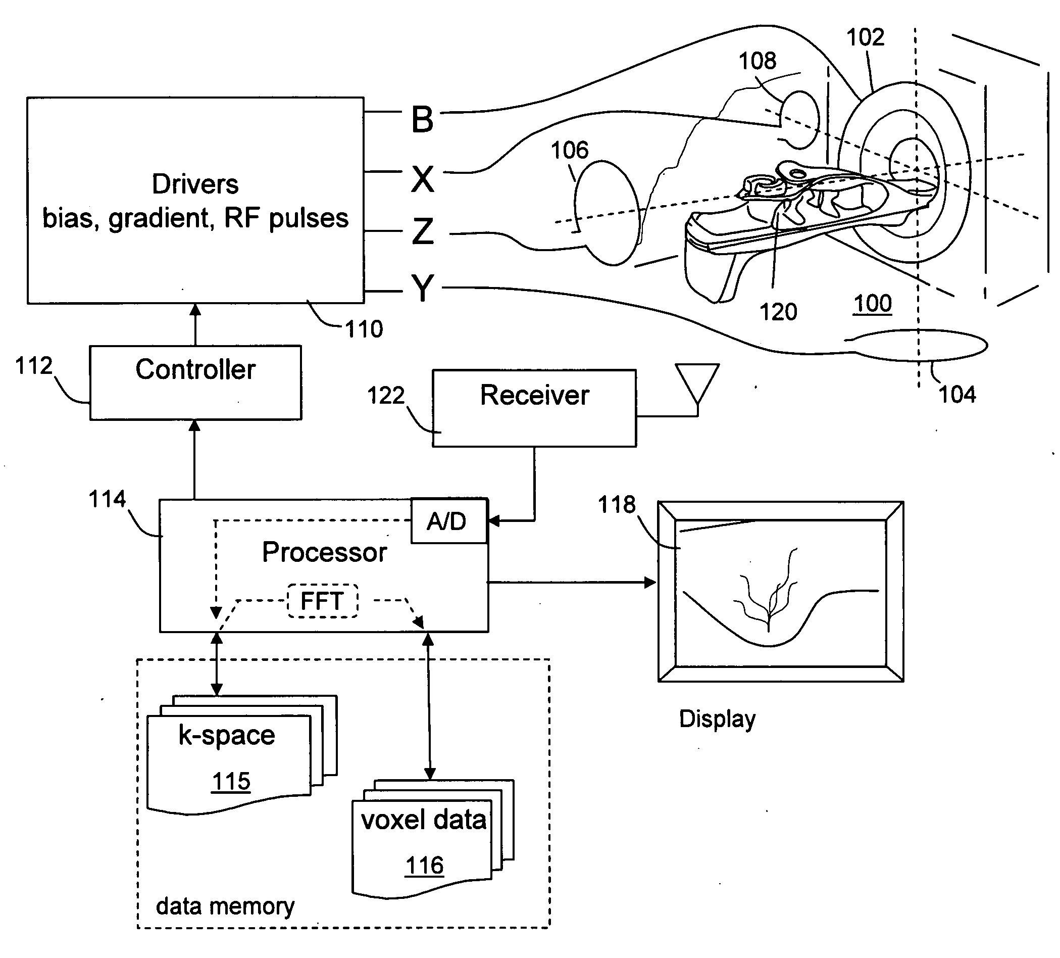
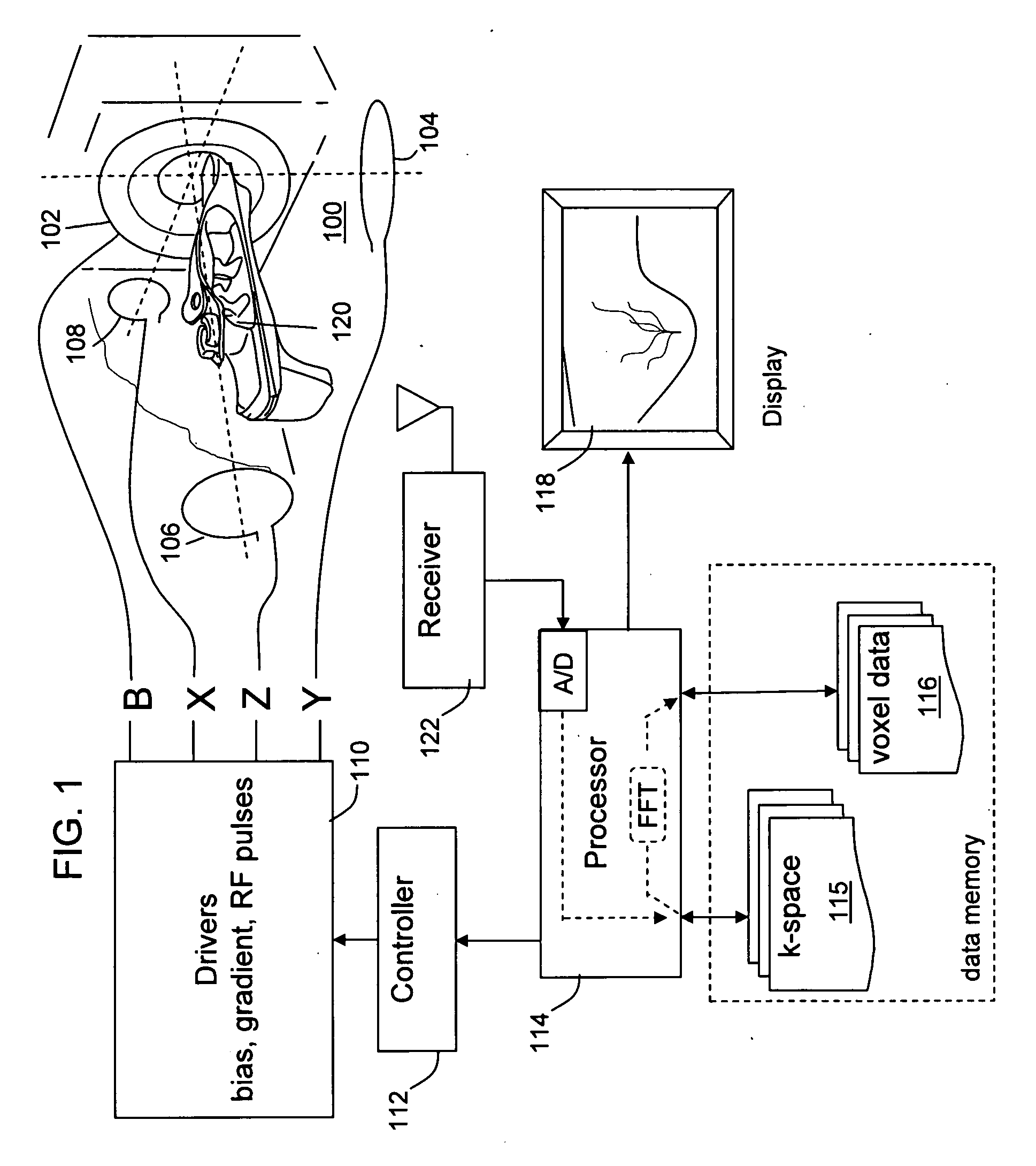
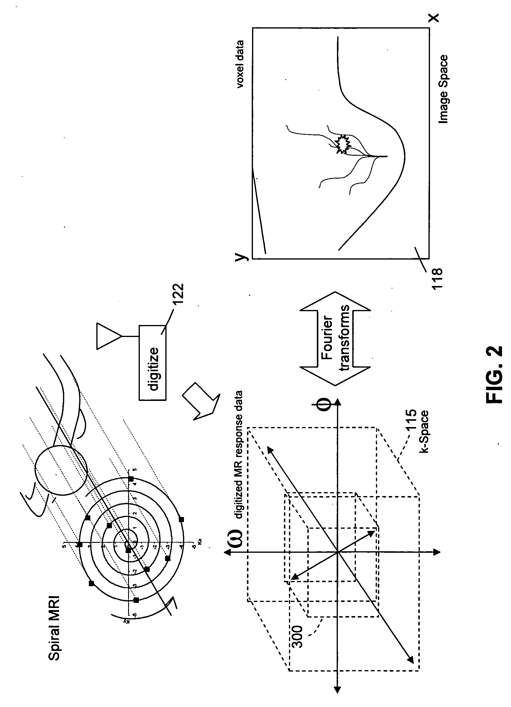
[0038]FIG. 1 shows generally the elements of a nuclear magnetic resonance (NMR or MRI) imaging system. In one embodiment, the imaging system can be a breast imaging system operated with rotating off-resonance excitation at frequencies chosen to distinguish water-based tissues while limiting the response of fat-based tissues. The system is configured to collect nuclear magnetic resonance information in a sequence of excitation and sensing operations that occurs while gradient magnetic fields are adjusted. A sequence is executed comprising excitation and phase encoding RF pulses. Each excitation is followed after a delay by a sensing interval during which the responsive signal from the imaged tissue is received, digitized and the results are stored in a data memory wherein digitized values are organized to populate a matrix conventionally known as k-space. As the sequence is executed, more and more of the image data is collected until data characterizing the response of the full tissu...


PUM


Abstract

An MRI image from spiral trajectory scanning is arranged as complementary subsets of values in time-sampled k-space. These values are Fourier transformed to produce a spatial domain image. While holding the patient stationary, the contrast information is updated at the central portion of k-space, and the peripheral portion of k-space data can be filled during the whole image acquisition. The contrast information is combined with the peripheral portion of k-space (contributing to image resolution) to construct a full k-space data and to generate a spatial image. The technique is useful for providing short time interval sampling when analyzing the take-up and fade-away of a contrast agent over time.

Description

FIELD OF THE INVENTION[0001]The invention relates to nuclear magnetic resonance imaging. Complementary subsets of k-space data from different MRI data acquisitions are combined, and the combinations are Fourier transformed to produce spatial images. A subset of k-space data from a central volume of k-space can be collected repeatedly in each successive MRI acquisition. The central k-space subset is combined with data from a peripheral volume in k-space that is collected less frequently, or even only once. Fourier transforming the combinations produces multiple images with a short acquisition time.BACKGROUND[0002]Nuclear magnetic resonance imaging (NMR or MRI) relies on the relaxation properties of nuclei in imaged volumes of tissue, when subjected to a steady state magnetic biasing field, excited by radio frequency signals. The tissue is caused to produce responsive electromagnetic radiation at locations that are addressed by timed gradient magnetic fields. Volumetric image data is ...

Claims


Application Information

Patent Timeline
Patent Type & Authority Applications(United States)
IPC IPC(8): A61B5/055
CPCA61B5/055A61B5/4312A61B5/7257G01R33/56341G01R33/5601G01R33/5619G01R33/4824
Inventor HONG, XIAOLEHARMS, STEVEN E.JIANG, AIPING
Owner AURORA HEALTHCARE US CORP
Why Eureka
Try Eureka