DNA methylation markers based on epigenetic stem cell signatures in cancer

a technology of epigenetic stem cells and methylation markers, applied in the field of dna methylation markers based on epigenetic stem cell signatures in cancer, can solve the problems of raising the specter of aberrant or rogue cells, unable to obtain firm empirical evidence, and no tool is available to sufficiently predict or monitor the efficacy of neoadjuvant or adjuvant systemic chemotherapy. , to achieve the effect of poor outcom

Active Publication Date: 2010-07-08
UNIV OF SOUTHERN CALIFORNIA +1
View PDF0 Cites 17 Cited by
  • Summary
  • Abstract
  • Description
  • Claims
  • Application Information

AI Technical Summary

Benefits of technology

[0018]In yet additional embodiments, various stem or precursor cells are used to identify transcriptional repressor occupancy sites (e.g., by chromatin immunoprecipitation chip analysis) and status for not only polycomb repressive complex 2 (PRC2), but also for other repressors and repressor complexes (e.g., repressors of developmental genes) as well, and these ChIP-Chip targets are then used as a means of enrichment for cancer-specific DNA methylation markers as taught herein using the exemplary combination of embryonic stems cells and PRC2 targets. According to further aspects, therefore, the instant approach has substantial utility for various types of stem and precursor cells (ES cell, somatic stem cells, hematopoietic stem cells, leukemic stem cells, skin stem cells, intestinal stem cells, gonadal stem cells, brain stem cells, muscle stem cells (muscle myoblasts, etc.), mammary stem cells, neural stem cells (e.g., cerebellar granule neuron progenitors, etc.), etc), and for various stem- or precursor cell repressor complexes (e.g., such as those described in Table 1 of Sparmann & Lohuizen, Nature 6, 2006 (Nature Reviews Cancer, November 2006), incorporated herein by reference), and for various types of cancer, where the requirements are that the repressor occupancy sites / loci and corresponding occupancy status are defined / established, and a characteristic DNA methylation status (e.g., disorder-specific, cancer-specific, etc.) (e.g., DNA hypermethylation) is established at corresponding sites / loci in one or more cellular proliferative disorders or cancers of interest, or, in particular embodiments, characteristic lineage-specific, stage specific, etc., status in cells of a developmental lineage or stage of interest.
[0019]Yet additional aspects provide a method for the diagnosis or prognosis of ovarian cancer comprising: performing methylation analysis of genomic DNA of a subject tissue sample; and determining the methylation state of a HOX genomic DNA sequence relative to a control HOX genomic DNA sequence, wherein diagnosis or prognosis of ovarian cancer is provided. In particular embodiments, the HOX genomic DNA sequence is that of HOXA10 or HOXA11, and hypermethylation is used to provide the ovarian cancer related diagnosis or prognosis. In certain aspects, the HOX genomic DNA sequence is that of HOXA11, and hypermethylation is used to provide a ovarian cancer related prognosis of poor outcome. In particular embodiments, the diagnostic or prognosic marker is for at least one selected from the group consisting of: for stem cells that are unable to differentiate; for stem cell that are resistant to therapy; for residual tumor after cytoreductive surgery; for cancer stem cells; for mucinous cancer cases; for serous cancer cases; for endometrioid cancer cases; for clear cell cases; and for tumor distribution.
[0020]Further aspects provide a method for predicting the response to neoadjuvant and / or adjuvant chemotherapy in a solid tumor, comprising performing methylation analysis of genomic DNA of a subject tissue sample; and determining the methylation state of a NEUROD1 genomic DNA sequence relative to a control NEUROD1 genomic DNA sequence, wherein predicting the response to neoadjuvant and / or adjuvant chemotherapy in breast cancer is provided. Additional aspects provide a method for determining chemosensitivity in breast cancer, comprising: performing methylation analysis of genomic DNA of a subject tissue sample; and determining the methylation state of a NEUROD1 genomic DNA sequence relative to a control NEUROD1 genomic DNA sequence, wherein determining chemosensitivity in breast cancer is provided. In certain embodiments of these methods, NEUROD1 methylation is a chemosensitivity marker in estrogen receptor (ER) negative breast cancer. In particular aspects, methylation analysis is at least one of: methylation analysis in core breast cancer biopsies taken prior to preoperative chemotherapy with complete pathological response as the endpoint; and seroconversion of NEUROD1 methylation in serum DNA during adjuvant chemotherapy with survival as the endpoint. In particular implementations, the chemosensitivity is with respect to at least one of cyclophospamide, methotrexate, 5-fluorouracil, anthracycline, and combinations thereof.

Problems solved by technology

However, it has been challenging to obtain firm empirical evidence supporting a normal stem cell origin of cancer and this question remained open.
However, these therapeutic strategies inevitably involve the introduction of human cells that have been maintained, manipulated, and / or differentiated ex vivo to provide the desired precursor cells (e.g., somatic stem cells, etc.), raising the specter that aberrant or rogue cells (e.g., cancer cells or cells predisposed to cancer that may occur during such manipulations and differentiation protocols) may be administered along with desired cells.
To date, however, no tool is available to sufficiently predict or monitor efficacy of neoadjuvant or adjuvant systemic chemotherapy which is frequently applied in ER negative breast cancer.

Method used


Image

  • DNA methylation markers based on epigenetic stem cell signatures in cancer
  • DNA methylation markers based on epigenetic stem cell signatures in cancer
  • DNA methylation markers based on epigenetic stem cell signatures in cancer

Examples


example 1

Methods

Colorectal Cancer Methods

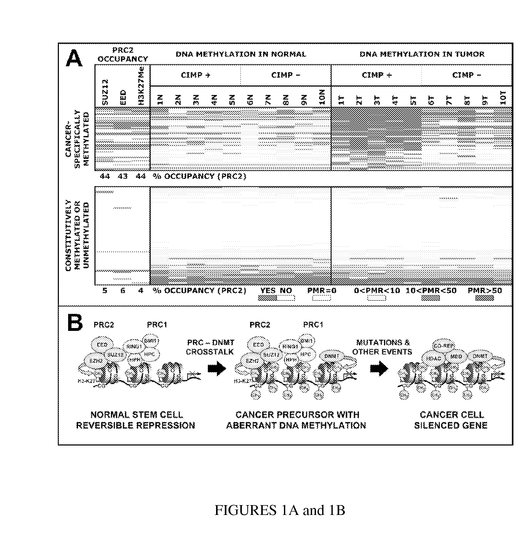
[0048]Colorectal cancer DNA Methylation Data and PRC2 Occupancy. The full methods for the colorectal cancer data have been published previously (D. J. Weisenberger et al., Nat Genet. 38:7, 2006; incorporated by reference herein in its entirety).

Methods Applicable to the Previously Unpublished Data for Ovarian Cancer, Breast Cancer, and CD34 Positive Hematopoietic Cells

Patients:

[0049]Hematopoietic related Patients. CD34 pos. cells isolated from stem cell apheresis collections from nine women were analyzed. The samples were collected during treatment at the Division of Hematology and Oncology, Innsbruck Medical University, Austria. All patients signed informed consent prior to apheresis.

[0050]Ovarian and breast related patients. Ovarian tissues from 40 patients and breast specimens from 30 patients were collected during surgery at the Department of Obstetrics and Gynecology of the Innsbruck Medical University, Austria in compliance with and approved by ...

example 2

Colorectal Cancer DNA Methylation Data and PRC2 Occupancy were Analyzed

[0059]Table 1 lists the 177 MethyLight™ reactions from Weisenberger et al. (2006) for which the PRC2 occupancy could be established from the data published in Lee et al. (2006). Of the 177 reactions, 164 (93%) are located within 1 kb of the transcription start site. Of the PRC2 targets, 95% are located within 1 kb of the transcription start site. See Table 5 herein below for primer and probe details.

TABLE 1Colorectal Cancer DNA Methylation Data and PRC2 OccupancyDNA METHYLATIONPRC2 OCCUPANCYMEANMEANHGNCREACTIONPRC2PMRPMRPMR(T) −SYMBOLIDSUZ12EEDH3K27MeTOTAL(N)(T)PMR(N)CANCER-SPECIFICALLY METHYLATED GENESGATA5HB-326YESYESYES335514479.00SFRP5HB-282YESYESYES33446443.45IGF2HB-319YESYESNO22368366.11TWIST1HB-047NOYESYES29294284.89EBF3HB-229YESNOYES213287273.78HIC1HB-168NOYESYES290356266.16SFRP2HB-280NONONO07187179.71SFRP1HB-201YESYESYES329177148.52NEUROD2HB-260YESYESYES326173147.11SCGB3A1HB-194NONONO07143135.44RUNX3HB-1...

example 3

Ovarian Cancer DNA Methylation Data and Stem Cell PRC2 Occupancy were Analyzed

[0060]Table 2 lists DNA methylation values (PMR) of 35 genes analyzed in 18 normal ovaries and 22 ovarian cancers. These genes were selected for their potential utility as cancer-specific DNA methylation markers without prior knowledge of their PRC2 occupancy status. P-values of genes that demonstrate significant higher DNA methylation levels (Mann Whitney U test) in cancer compared to normal ovaries are shaded and referred as to “cancer genes”. Applicants defined “Stem cell genes” as genes which are occupied with at least two of the three components (SUZ12, EED and H3K27me3) in human embryonic stem cells. Nine genes demonstrated higher frequencies of densely methylated alleles (as reflected in the listed values for PMR) in cancer tissues compared to normal ovaries. 56% ( 5 / 9) of these “cancer genes” were “stem cell genes”, whereas only 15% ( 4 / 26) of the “non-cancer genes” were “stem cell genes” (P=0.03)....


PUM


Abstract

In particular aspects, stem-cell polycomb group (PcG) targets are more likely to have cancer-specific promoter DNA methylation than non-targets, indicating a stem-cell origin of cancer, where reversible gene repression is replaced by permanent silencing, locking the cell into a perpetual state of self-renewal and predisposition to subsequent malignant transformation. Exemplary aspects provide methods for identifying preferred DNA methylation markers for a cellular proliferative disorder and/or cancer and markers for developmental lineages and/or stages, based on identifying PcG protein or PcG repressive complex genomic target loci within a precursor cell (e.g., stem or progenitor cell) population, and determining, in cells of the proliferative disorder and/or cancer or cell of the particular developmental lineages and/or stages, a characteristic methylation status of the PcG target loci. Additional aspects provide methods for validating and/or monitoring a precursor cell (e.g., stem cell) population. Diagnostic and prognostic methods for ovarian and breast cancer are provided.

Description

CROSS-REFERENCE TO RELATED APPLICATIONS[0001]This application claims the benefit of priority to U.S. Provisional Patent Application Ser. Nos. 60 / 877,530, filed 27 Dec. 2006 and entitled “DNA METHYLATION MARKERS BASED ON EPIGENETIC STEM CELL SIGNATURE IN CANCER,” and 60 / 882,948, filed 31 Dec. 2006 and of same title, both of which are incorporated herein by reference in their entirety.STATEMENT REGARDING FEDERALLY-SPONSORED RESEARCH[0002]Particular aspects of this inventive subject matter were supported by National Institutes of Health (NIH) grant R01 CA075090, and the Untied States government may, therefore, have certain rights to these aspects of the invention.FIELD OF THE INVENTION[0003]Particular aspects relate generally to markers (e.g., diagnostic and / or prognostic DNA methylation markers) for cellular proliferative disorders and / or cancer and markers or for developmental lineages and / or stages, and to precursor cells (e.g., embryonic stem (ES) cells, somatic stem cells, cancer ...

Claims


Application Information

Patent Timeline
Patent Type & Authority Applications(United States)
IPC IPC(8): A61K35/12C12Q1/68A61P35/04
CPCC12Q1/6886C12Q2600/106C12Q2600/158C12Q2600/154C12Q2600/118A61P35/04
Inventor LAIRD, PETER W.WIDSCHWENDTER, MARTIN
Owner UNIV OF SOUTHERN CALIFORNIA
Why Eureka
Try Eureka