System for Cardiac Ultrasound Image Acquisition

a technology of cardiac ultrasound and image acquisition, applied in the field of ultrasound medical imaging system, can solve the problems of limited gating capability for studying particular cardiac functions, inability to effectively scan for optimal imaging, and failure of known systems to comprehensively perform cardiac function tracking of maximum size and volume of ventricle chambers

Inactive Publication Date: 2010-08-19
SIEMENS HEATHCARE GMBH
View PDF22 Cites 15 Cited by
  • Summary
  • Abstract
  • Description
  • Claims
  • Application Information

AI Technical Summary

Benefits of technology

[0005]An ultrasound medical imaging system automatically determines image resolution, scanning frequency and acquisition speed in response to data indicating a clinical application and cardiac function signals including hemodynamic (blood pressure) signals and vital sign (such as SPO2, respiration, NIBP) signals used to trigger and synchronize image scanning and data acquisition. An ultrasound medical imaging system adaptively acquires anatomical images using an ultrasound image acquisition device. The ultrasound image acquisition device initiates acquisition of anatomical images of a portion of patient anatomy in response to a heart rate related synchronization signal. The ultrasound image acquisition device includes multiple ultrasound transdu

Problems solved by technology

However known ultrasound image scanning and acquisition systems typically employ a fixed time interval between frames and fail to effectively scan for optimum imaging results for real time clinical monitoring and diagnosis.
Known systems employ surface ECG signals for image gating to avoid cardiac contraction noise, for example, but offer limited gating capability for studying particular cardiac functions.
However, known systems fail to comprehensively perform cardiac function tracking of maximum size and volume of ventricle chambers, for example, in the presence o

Method used


Image

  • System for Cardiac Ultrasound Image Acquisition
  • System for Cardiac Ultrasound Image Acquisition
  • System for Cardiac Ultrasound Image Acquisition

Examples


Embodiment Construction

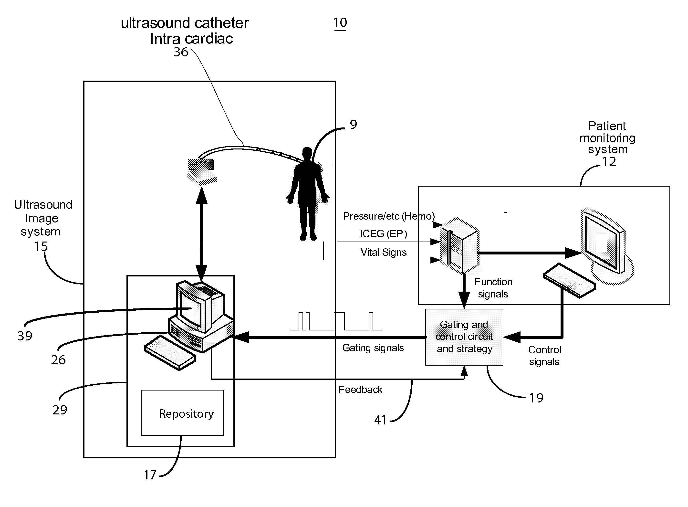
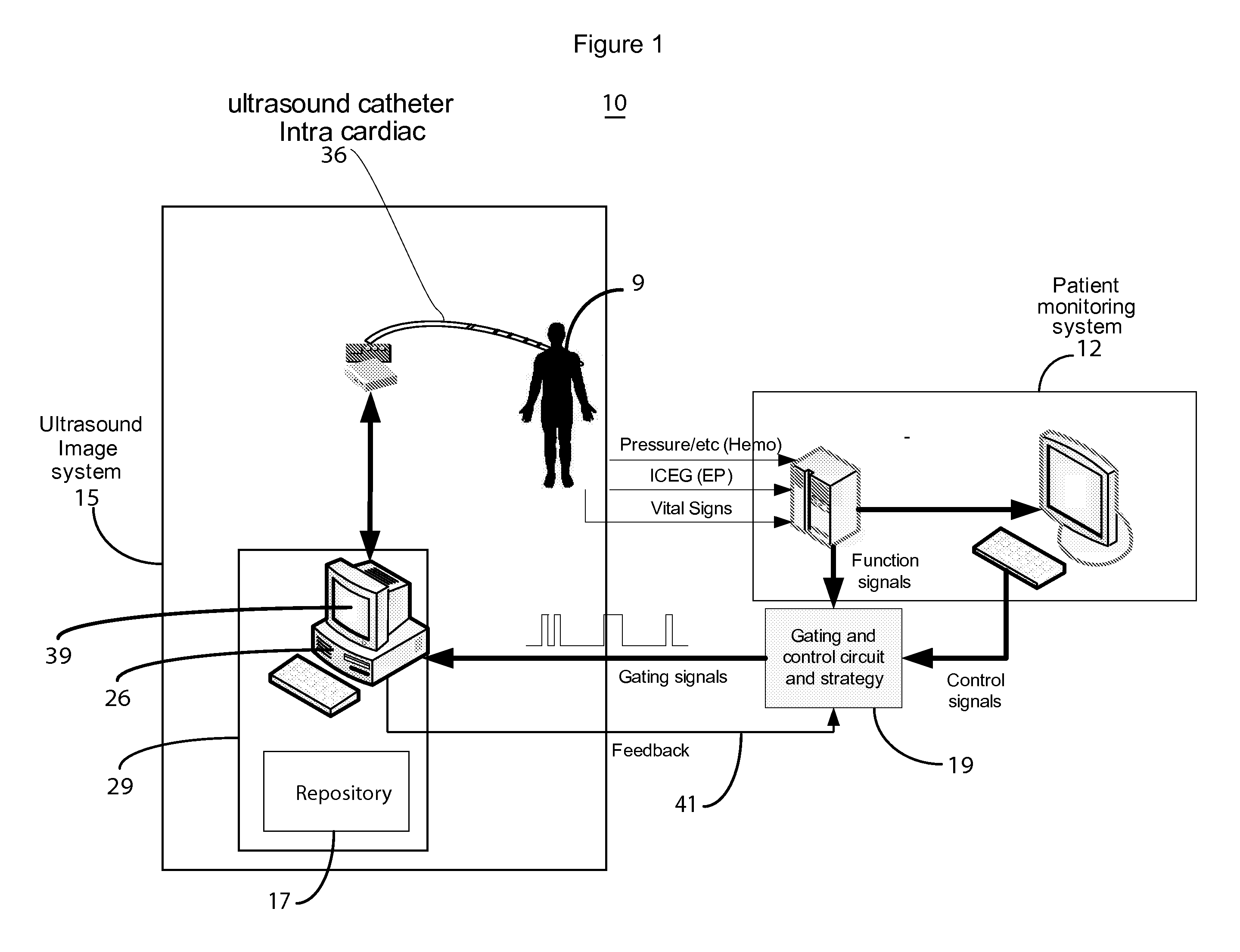
[0015]A system improves medical imaging using non-uniform and nonlinear cardiac functional signals such as hemodynamic signals (invasive blood pressure, non-invasive blood pressure, blood flow speed), electrophysiological signals (surface ECG, intra-cardiac electrograms, both unipolar and bipolar signals), and vital signs signals (SPO2, respiration), to trigger and synchronize image scanning and data acquisition. The image resolution, scanning frequency and acquisition speed of the ultrasound image system is automatically determined in response to cardiac functions and a clinical application. The system uses ultrasound imaging for better quantitative and qualitative diagnosis and characterization of cardiac function and patient health status.

[0016]The system image gating and synchronization function is advantageously adaptively dynamically configured to use one or a combination of signals for different kinds of clinical applications and procedures. The combination of signals include...


PUM


Abstract

An ultrasound image acquisition device initiates acquisition of anatomical images of a portion of patient anatomy in response to a heart rate related synchronization signal. The ultrasound image acquisition device includes multiple ultrasound transducers for generating sound waves. The ultrasound transducers are arranged in different transducer groups oriented to enable acquisition of different ultrasound imaging information used in generating a single composite ultrasound image. A synchronization processor derives the heart rate related synchronization signal from a patient cardiac function blood flow related parameter. The synchronization signal enables adaptive activation of a particular group of the different transducer groups for acquisition of ultrasound imaging information used in generating the single composite ultrasound image. A display processor presents the single composite ultrasound image, acquired by the ultrasound image acquisition device, to a user on a reproduction device.

Description

[0001]This is a non-provisional application of provisional application Ser. No. 61 / 153,067 filed Feb. 17, 2009, by H. Zhang.FIELD OF THE INVENTION[0002]This invention concerns an ultrasound medical imaging system for adaptively acquiring anatomical images involving activating different transducer groups for acquisition of ultrasound imaging information presented in a composite ultrasound image.BACKGROUND OF THE INVENTION[0003]Ultrasound medical imaging is used for imaging the heart and surrounding intra-thoracic structures. However known ultrasound image scanning and acquisition systems typically employ a fixed time interval between frames and fail to effectively scan for optimum imaging results for real time clinical monitoring and diagnosis. Known systems employ surface ECG signals for image gating to avoid cardiac contraction noise, for example, but offer limited gating capability for studying particular cardiac functions. Continuous image scanning and acquisition, such as intra-...

Claims


Application Information

Patent Timeline
IPC IPC(8): A61B8/14A61B5/0402A61B8/00
CPCA61B5/0205A61B6/466A61B8/08A61B8/5238A61B8/543A61B8/4472A61B8/0883
Inventor ZHANG, HONGXUAN
Owner SIEMENS HEATHCARE GMBH
Why Eureka
Try Eureka