Device for collecting samples

a technology for collecting samples and uterine secretions, which is applied in the field of collecting cytological, bacteriological and uterine secretions samples, can solve the problems of difficult impossibility of collecting clean materials, and difficult collection of pure materials in animal species, etc., to achieve the effect of improving the penetration of the uterine cervix and small diameter

Inactive Publication Date: 2010-09-16
EESTI MAAULIKOOL ESTONIAN UNIV OF LIFE SCI
View PDF8 Cites 8 Cited by
  • Summary
  • Abstract
  • Description
  • Claims
  • Application Information

AI Technical Summary

Benefits of technology

[0011]The Cytobrush guiding tube cap has a smaller diameter than the Cytobrush guiding tube to improve uterine cervical penetration. Transition from the guiding tube cap to the guiding tube is smooth. The Cytobrush ball, together with the guiding tube cap, forms the smooth transition to the guiding tube.
[0012]To avoid the contamination of the Cytobrush, the guiding tube cap is equipped with an inward-facing cone so that the Cytobrush ball fits tightly inside the guiding tube cap.
[0013]The inner diameter of the Cytobrush guiding tube cap is designed so that only the Cytobrush fits into it, but not the Cytobrush fixating rod. Such a design makes it possible to advance the Cytobrush from the guiding tube cap an optimal distance and avoid the Cytobrush penetrating too far into the uterus. Such a design guarantees sample collection precisely from the desired area.

Problems solved by technology

The collection of pure material is more difficult in animal species (e.g. cows) whose uterine cervix is difficult to penetrate due to the anatomical build and / or the physiological status of the animal.
The use of an unsuitable device to penetrate the uterine cervix may damage it, creating favourable conditions for inflammation; and excessive manipulation of the device (by moving it back and forth) creates the risk of its contamination, which leads to the impossibility of collecting clean material, and this may affect the analysis result and diagnostic precision.
Another important problem is the collecting of clean material from animals with a high body weight.
Even the smallest movement of an animal with a high body weight may cause the contamination of the device during sample collection.
The shortcomings of this device when penetrating the uterine cervix are the possibility of uterine cervical damage, which creates favourable conditions for inflammation; and the risk of device contamination when it is extensively manipulated (moved back and forth), which makes it impossible to collect a clean sample and may influence the analysis result.

Method used


Image

  • Device for collecting samples
  • Device for collecting samples
  • Device for collecting samples

Examples


Embodiment Construction

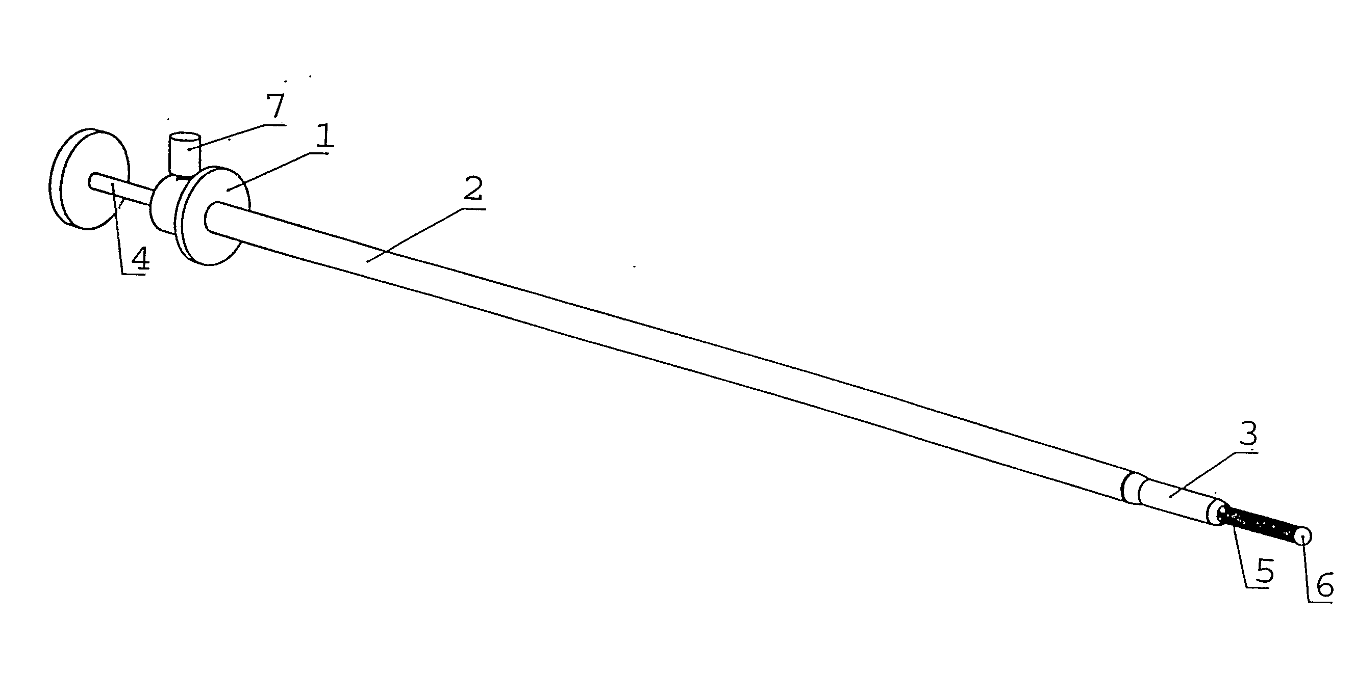
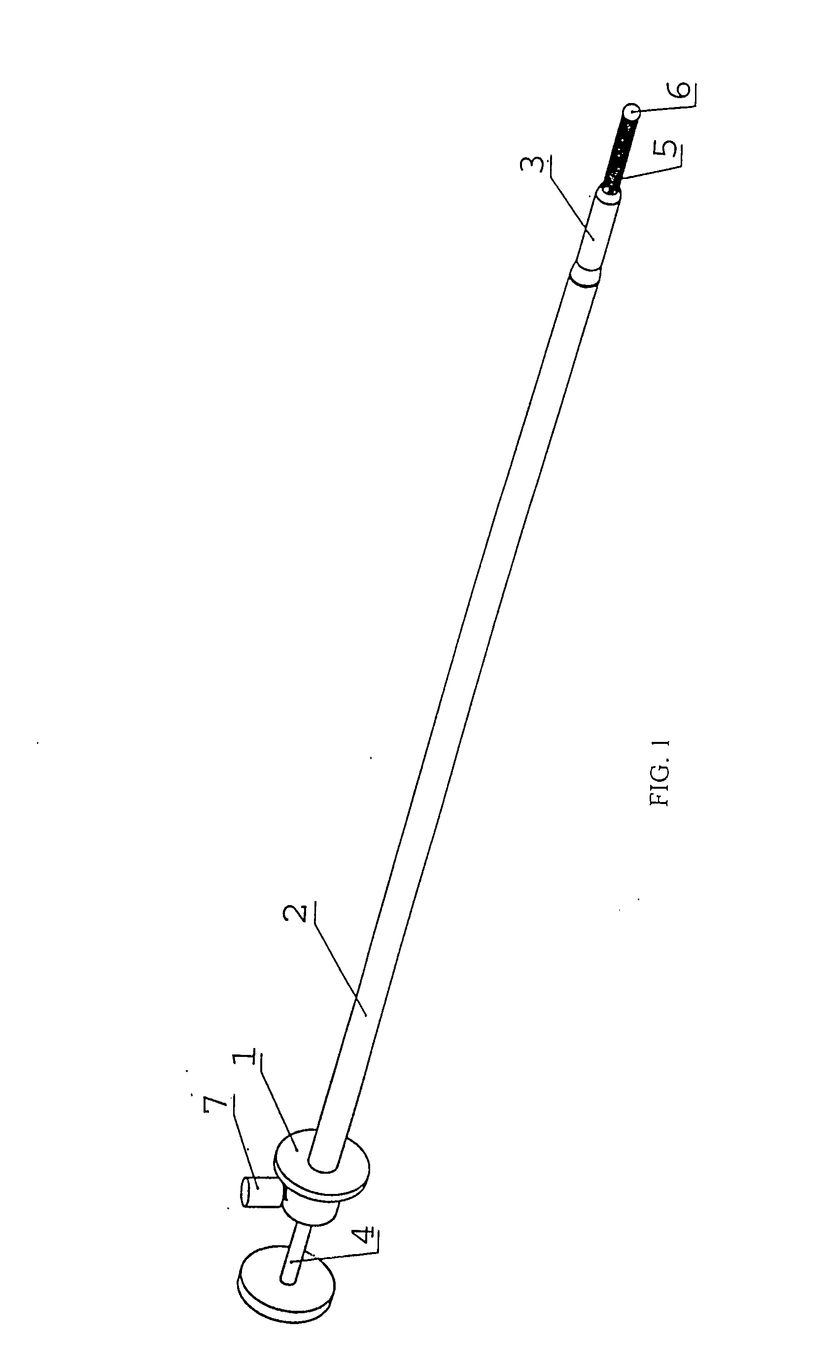
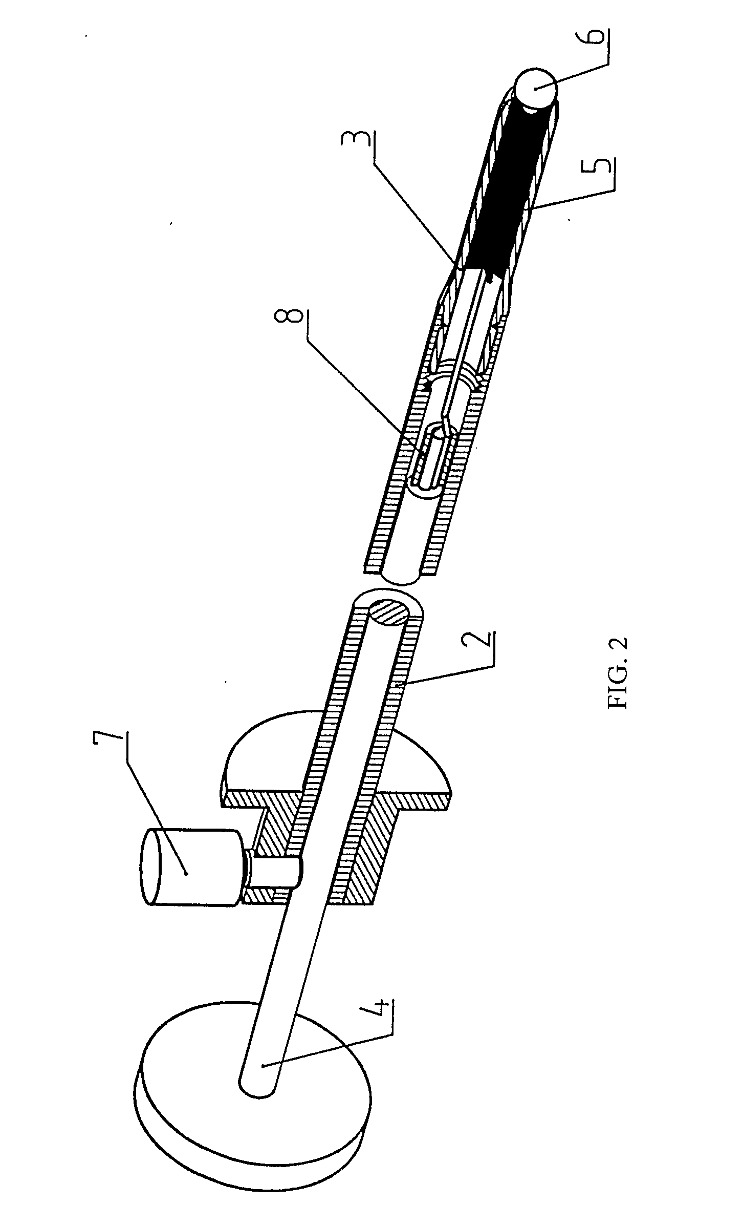
[0016]Before sample collection, the device 1 was sterilized. Before the application of the device, the device 1 was set in its working position by unfastening the rod-fixating screw 7. The Cytobrush was retracted into the guiding tube cap 3 with the help of the device's rod 4. The Cytobrush was fixated with the rod-fixating screw 7 and the device 1 was then introduced into the uterus via the vagina and uterine cervix. When the device 1 was introduced into the desired region, the rod-fixating screw 7 was unfastened and the rod was pushed into the guiding tube 2, as a result of which the Cytobrush advanced into the uterus, where it contacted with the endometrium. By rotating the rod 4 multiple times around its axle, endometrium material was collected on the Cytobrush 4. Then the Cytobrush was retracted into the guiding cap 3 with the help of rod 4. The rod 4 was fixated with the help of the rod-fixating screw 7 and the device 1 was taken out of the uterus and the animal. The fixation ...


PUM


Abstract

The present invention belongs to the veterinary field and will be applied to collect cytological, bacteriological and uterine secretion samples primarily from the uterine endometrium with the aim of detecting inflammations, including subclinical endometritis. The invention relates to a sampling device for obtaining cytological, bacteriological and smear samples from uterus of animals whose uterine cervix is difficult to penetrate. The device enables to obtain clean diagnostic material, which is important in obtaining high diagnostic accuracy. The device comprises a guiding tube and a rod extending through the guiding tube with a brush attached, and containing in addition a guiding tube cap that can be attached to a guiding tube with an inwards-facing cone, a Cytobrush ball and a rod-fixating screw located on the rear end of the guiding tube.

Description

TECHNICAL FIELD OF INVENTION[0001]The invention belongs to the veterinary field and will be applied to collect cytological, bacteriological and uterine secretion samples primarily from the uterine endometrium with the aim of detecting inflammations, including subclinical endometritis.BACKGROUND OF THE INVENTION[0002]To detect the inflammations, the presence and counts of white blood cells and bacteria in cytological and bacteriological material and presence and quantity of inflammation markers in uterine secretion are determined. On the basis of the presence and count of white blood cells and bacteria and inflammation markers it is possible to diagnose uterine inflammations.[0003]The maximum diagnostic precision is guaranteed by the cleanness of the material. The collection of pure material is more difficult in animal species (e.g. cows) whose uterine cervix is difficult to penetrate due to the anatomical build and / or the physiological status of the animal. The use of an unsuitable ...

Claims


Application Information

Patent Timeline
Patent Type & Authority Applications(United States)
IPC IPC(8): A61B10/02
CPCA61B10/0291A61B2017/00336A61B2010/0216
Inventor VALDMANN, ANDRESVALDMANN, MERLE
Owner EESTI MAAULIKOOL ESTONIAN UNIV OF LIFE SCI
Why Eureka
Try Eureka